Effects of systemic ozone administration on the fresh extraction sockets healing: a histomorphometric and immunohistochemical study in rats
DOI:
https://doi.org/10.1590/1678-7757-2023-0412Keywords:
Ozone therapy, Bone healing, Animal research, RatsAbstract
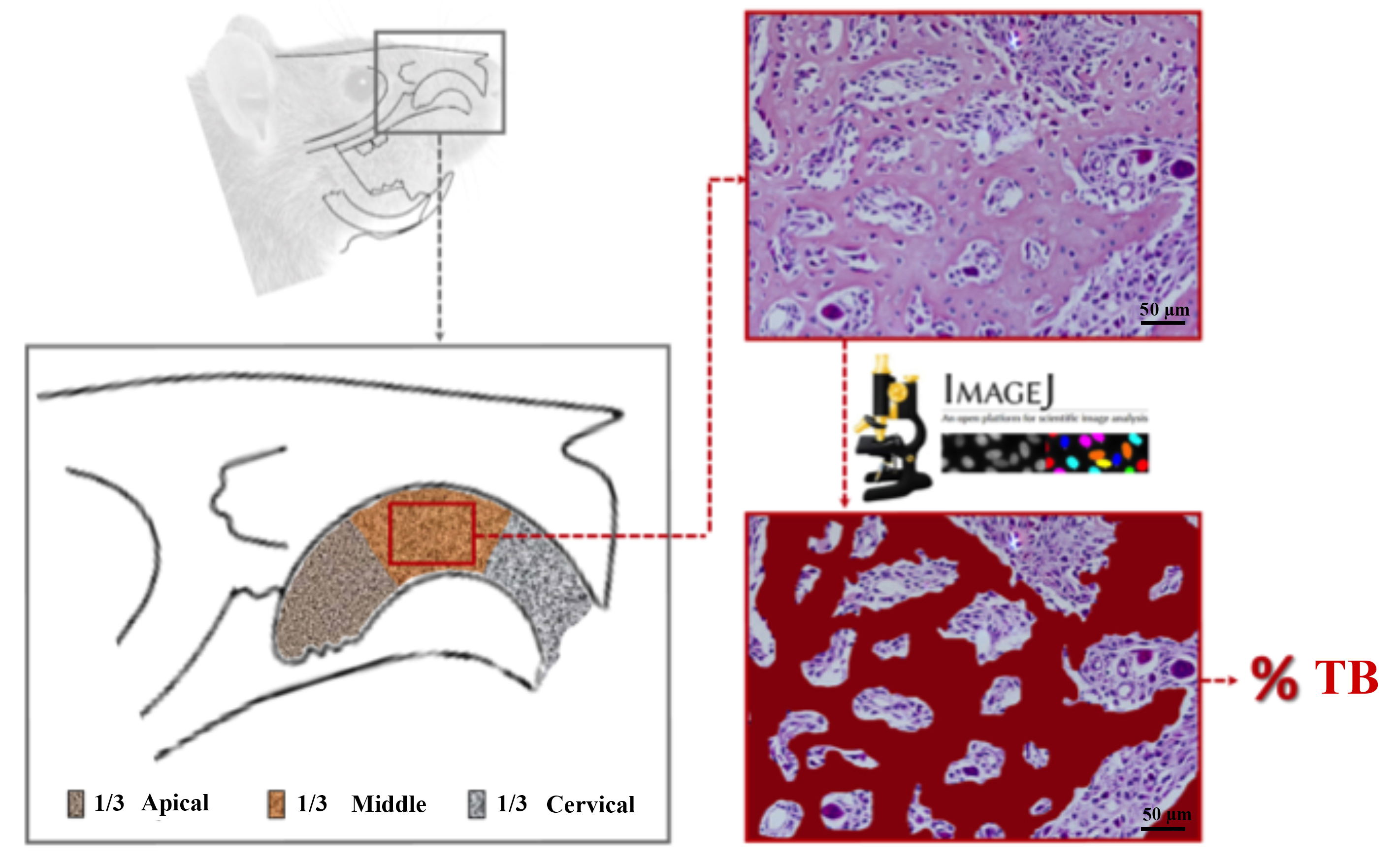
Studies have highlighted numerous benefits of ozone therapy in the field of medicine and dentistry, including its antimicrobial efficacy against various pathogenic microorganisms, its ability to modulate the immune system effectively, reduce inflammation, prevent hypoxia, and support tissue regeneration. However, its effects on dental extraction healing remain to be elucidated. Objective: Therefore, this study aimed to evaluate the effects of systemically administered ozone (O3) at different doses in the healing of dental extraction sockets in rats. Methodology: To this end, 72 Wistar rats were randomly divided into four groups after extraction of the right upper central incisor: Group C – control, no systemic treatment; Group OZ0.3 – animals received a single dose of 0.3 mg/kg O3; Group OZ0.7 – a single dose of 0.7 mg/kg O3; and Group OZ1.0 – a single dose of 1.0 mg/kg O3, intraperitoneally. In total, six animals from each group were euthanized at 7, 14, and 21 days after the commencement of treatment. Bone samples were harvested and further analyzed by descriptive histology, histomorphometry, and immunohistochemistry for osteocalcin (OCN) and tartrate-resistant acid phosphatase (TRAP) protein expression. Results: All applied doses of O3 were shown to increase the percentage of bone tissue (PBT) after 21 days compared to group C. After 14 days, the OZ0.7 and OZ1.0 groups showed significantly higher PBT when compared to group C. The OZ1.0 group presented the most beneficial results regarding PBT among groups, which denotes a dose-dependent response. OCN immunostaining was higher in all groups at 21 days. However, after seven and 14 days, the OZ1.0 group showed a significant increase in OCN immunostaining compared to C group. No differences in TRAP+ osteoclasts were found between groups and time points. Conclusion: Therefore, O3 therapy at higher doses might be beneficial for bone repair of the alveolar socket following tooth extraction.
Downloads
References
Barbe A, Mikhailenko S, Starikova E, Tyuterev V. High resolution infrared spectroscopy in support of ozone atmospheric monitoring and validation of the potential energy function. Molecules. 2022;27(3):911. doi: 10.3390/molecules27030911
Nogales CG, Ferrari PH, Kantorovich EO, Lage-Marques JL. Ozone therapy in medicine and dentistry. J Contemp Dent Pract. 2008;9(4):75-84
Baysan A, Lynch E. The use of ozone in dentistry and medicine. Prim Dent Care. 2005;12(2):47-52. doi: 10.1308/1355761053695158
El Meligy OA, Elemam NM, Talaat IM. Ozone therapy in medicine and dentistry: a review of the literature. Dent J (Basel). 2023;11(8):187. doi: 10.3390/dj11080187
Talukdar A, Langthasa M, Talukdar P, Barman I. Ozone therapy-boon to dentistry and medicine. Int J Prev Clin Dent Res. 2015;2(1):59-66.
Filipovic-Zore I, Divic Z, Duski R, Gnjatovic N, Galic N, Prebeg D. Impact of ozone on healing after alveolectomy of impacted lower third molars. Saudi Med J. 2011;32(6):642-44.
Bocci V, Zanardi I, Travagli V. Oxygen/ozone as a medical gas mixture: a critical evaluation of the various methods clarifies positive and negative aspects. Med Gas Res. 2011;1(1):6. doi: 10.1186/2045-9912-1-6
Cardoso MG, Oliveira LD, Koga-Ito CY, Jorge AO. Effectiveness of ozonated water on Candida albicans, Enterococcus faecalis, and endotoxins in root canals. Oral Surg Oral Med Oral Pathol Oral Radiol Endod. 2008;105(3):e85-91. doi: 10.1016/j.tripleo.2007.10.006
Clavo B, Perez JL, Lopez L, Suarez G, Lloret M, Rodriguez V, et al. Ozone therapy for tumor oxygenation: a pilot study. Evid Based Complement Alternat Med. 2004;1(1):93-8. doi: 10.1093/ecam/neh009
Cespedes-Suarez J, Martin-Serrano Y, Carballosa-Peña MR, Dager-Carballosa DR. The immune response behavior in HIV-AIDS patients treated with Ozone therapy for two years. J Ozone Ther. 2018;3(2):111458. doi: 10.7203/jo3t.2.3.2018.11458
Setyo Budi D, Fahmi Rofananda I, Reza Pratama N, Sutanto H, Sukma Hariftyani A, Ratna Desita S, et al. Ozone as an adjuvant therapy for COVID-19: a systematic review and meta-analysis. Int Immunopharmacol. 2022;110:109014. doi: 10.1016/j.intimp.2022.109014
Erdemci F, Gunaydin Y, Sencimen M, Bassorgun I, Ozler M, Oter S, et al. Histomorphometric evaluation of the effect of systemic and topical ozone on alveolar bone healing following tooth extraction in rats. Int J Oral Maxillofac Surg. 2014;43(6):777-83. doi:10.1016/j.ijom.2013.12.007
Srikanth A, Sathish M, Sri Harsha AV. Application of ozone in the treatment of periodontal disease. J Pharm Bioallied Sci. 2013;5(Suppl1):S89-94. doi: 10.4103/0975-7406.113304
Stubinger S, Sader R, Filippi A. The use of ozone in dentistry and maxillofacial surgery: a review. Quintessence Int. 2006;37(5):353-9.
Lima TJ Neto , Delanora LA, Simon ME, Ribeiro KH, Matsumoto MA, Louzada MJ, et al. Ozone improved bone dynamic of female rats using zoledronate. Tissue Eng Part C Methods. 2024 Jan;30(1):1-14. doi: 10.1089/ten.TEC.2023.0159
Cosola S, Giammarinaro E, Genovesi AM, Pisante R, Poli G, Covani U, et al. A short-term study of the effects of ozone irrigation in an orthodontic population with fixed appliances. Eur J Paediatr Dent. 2019;20(1):15-18. doi: 10.23804/ejpd.2019.20.01.03
Hodson N, Dunne SM. Using ozone to treat dental caries. J Esthet Restor Dent. 2007;19(6):303-5. doi: 10.1111/j.1708-8240.2007.00127.x
Saini R. Ozone therapy in dentistry: a strategic review. J Nat Sci Biol Med. 2011;2(2):151-3. doi: 10.4103/0976-9668.92318
Kollmuss M, Kist S, Obermeier K, Pelka AK, Hickel R, Huth KC. Antimicrobial effect of gaseous and aqueous ozone on caries pathogen microorganisms grown in biofilms. Am J Dent. 2014;27(3):134-8.
Huth KC, Quirling M, Maier S, Kamereck K, Alkhayer M, Paschos E, et al. Effectiveness of ozone against endodontopathogenic microorganisms in a root canal biofilm model. Int Endod J. 2009;42(1):3-13. doi: 10.1111/j.1365-2591.2008.01460.x
Nagayoshi M, Kitamura C, Fukuizumi T, Nishihara T, Terashita M. Antimicrobial effect of ozonated water on bacteria invading dentinal tubules. J Endod. 2004;30(11):778-81. doi: 10.1097/00004770-200411000-00007
Randi CJ, Heiderich CM, Serrano RV, Morimoto S, Moraes LO, Campos L, et al. Use of ozone therapy in implant dentistry: a systematic review. Oral Maxillofac Surg. 2023;28(1):39-49. doi: 10.1007/s10006-023-01149-3
Alsakr A, Gufran K, Alqahtani AS, Alasqah M, Alnufaiy B, Alzahrani HG, et al. Ozone therapy as an adjuvant in the treatment of periodontitis. J Clin Med. 2023;12(22):7078. doi: 10.3390/jcm12227078
Mehrotra R, Gupta S, Siddiqui ZR, Chandra D, Ikbal SA. Clinical efficacy of ozonated water and photodynamic therapy in non-surgical management of chronic periodontitis: A clinico- microbial study. Photodyn Ther. 2023;44:103749. doi: 10.1016/j.pdpdt.2023.103749
Scribante A, Gallo S, Pascadopoli M, Frani M, Butera A. Ozonized gels vs chlorhexidine in non-surgical periodontal treatment: A randomized clinical trial. Oral Dis. Forthcoming 2023. doi: 10.1111/odi.14829
Tatuskar PV, Karmakar S, Walavalkar NN, Vandana KL. Effect of ozone irrigation and powered toothbrushing on dental plaque, gingival inflammation, and microbial status in institutionalized mentally challenged individuals: a double-blinded, randomized, controlled clinical trial. J Indian Soc Periodontol. 2023;27(3):315-9. doi: 10.4103/jisp.jisp_69_22
Suvan J, Leira Y, Moreno Sancho FM, Graziani F, Derks J, Tomasi C. Subgingival instrumentation for treatment of periodontitis. A systematic review. J Clin Periodontol. 2020;47 Suppl 22:155-175. doi: 10.1111/jcpe.13245
Colombo M, Gallo S, Garofoli A, Poggio C, Arciola CR, Scribante A. Ozone gel in chronic periodontal disease: a randomized clinical trial on the anti-inflammatory effects of ozone application. Biology (Basel). 2021;10(7):625. doi: 10.3390/biology10070625
Sen S, Sen S. Ozone therapy a new vista in dentistry: integrated review. Med Gas Res. 2020;10(4):189-92. doi: 10.4103/2045-9912.304226
Chandrasekhar T, Ratnaditya A, Kandregula CR, GM N. Ozone therapy: applications in preventive dentistry. J Res Adv Dent. 2015;4(1):1036.
Materni A, Pasquale C, Longo E, Frosecchi M, Benedicenti S, Bozzo M, et al. Prevention of dry socket with ozone oil-based gel after inferior third molar extraction: a double-blind split-mouth randomized placebo-controlled clinical trial. Gels. 2023;9(4):289. doi: 10.3390/gels9040289
Percie du Sert N, Ahluwalia A, Alam S, Avey MT, Baker M, Browne WJ, et al. Reporting animal research: explanation and elaboration for the ARRIVE guidelines 2.0. PLoS Biol. 2020;18(7):e3000411. doi: 10.1371/journal.pbio.3000411
Almeida CS, Sartoretto SC, Durte IM, Alves A, Barreto HV, Resende RFB, et al. In vivo evaluation of bovine xenograft associated with oxygen therapy in alveolar bone repair. J Oral Implantol. 2021;47(6):465-71. doi: 10.1563/aaid-joi-D-20-00110
Nogueira AV, Souza JA, Molon RS, Pereira ES, Aquino SG, Giannobile WV, et al. HMGB1 localization during experimental periodontitis. Mediators Inflamm. 2014;2014:816320. doi: 10.1155/2014/816320
Belluci MM, Molon RS, Rossa C Jr, Tetradis S, Giro G, Cerri PS, et al. Severe magnesium deficiency compromises systemic bone mineral density and aggravates inflammatory bone resorption. J Nutr Biochem. 2020;77:108301. doi: 10.1016/j.jnutbio.2019.108301
Santos PL, Molon RS, Queiroz TP, Okamoto R, Faloni AP, Gulinelli JL, et al. Evaluation of bone substitutes for treatment of peri-implant bone defects: biomechanical, histological, and immunohistochemical analyses in the rabbit tibia. J Periodontal Implant Sci. 2016;46(3):176-96. doi: 10.5051/jpis.2016.46.3.176
Ervolino E, Statkievicz C, Toro LF, Mello-Neto JM, Cavazana TP, Issa JP, et al. Antimicrobial photodynamic therapy improves the alveolar repair process and prevents the occurrence of osteonecrosis of the jaws after tooth extraction in senile rats treated with zoledronate. Bone. 2019;120:101-13. doi: 10.1016/j.bone.2018.10.014
Bocci V, Di Paolo N. Oxygen-ozone therapy in medicine: an update. Blood Purif. 2009;28(4):373-6. doi: 10.1159/000236365
Masan J, Sramka M, Rabarova D. The possibilities of using the effects of ozone therapy in neurology. Neuro Endocrinol Lett. 2021;42(1):13-21.
Kazancioglu HO, Ezirganli S, Aydin MS. Effects of laser and ozone therapies on bone healing in the calvarial defects. J Craniofac Surg. 2013;24(6):2141-6. doi: 10.1097/SCS.0b013e3182a244ae
Ozdemir H, Toker H, Balci H, Ozer H. Effect of ozone therapy on autogenous bone graft healing in calvarial defects: a histologic and histometric study in rats. J Periodontal Res. 2013;48(6):722-6. doi: 10.1111/jre.12060
Seidler V, Linetskiy I, Hubalkova H, Stankova H, Smucler R, Mazanek J. Ozone and its usage in general medicine and dentistry. a review article. Prague Med Rep. 2008;109(1):5-13.
Saglam E, Alinca SB, Celik TZ, Hacisalihoglu UP, Dogan MA. Evaluation of the effect of topical and systemic ozone application in periodontitis: an experimental study in rats. J Appl Oral Sci. 2020;28:e20190140. doi: 10.1590/1678-7757-2019-0140

Downloads
Published
Issue
Section
License
Copyright (c) 2024 Journal of Applied Oral Science

This work is licensed under a Creative Commons Attribution 4.0 International License.
Todo o conteúdo do periódico, exceto onde está identificado, está licenciado sob uma Licença Creative Commons do tipo atribuição CC-BY.

