
Performance of obturation techniques in anatomical irregularities located at different thirds of the root canal system
DOI:
https://doi.org/10.1590/1678-7757-2023-0440Keywords:
Anatomical irregularities, Gutta-percha filled area, Micro-CT-based tooth replica, Obturation techniques, Root canal anatomyAbstract
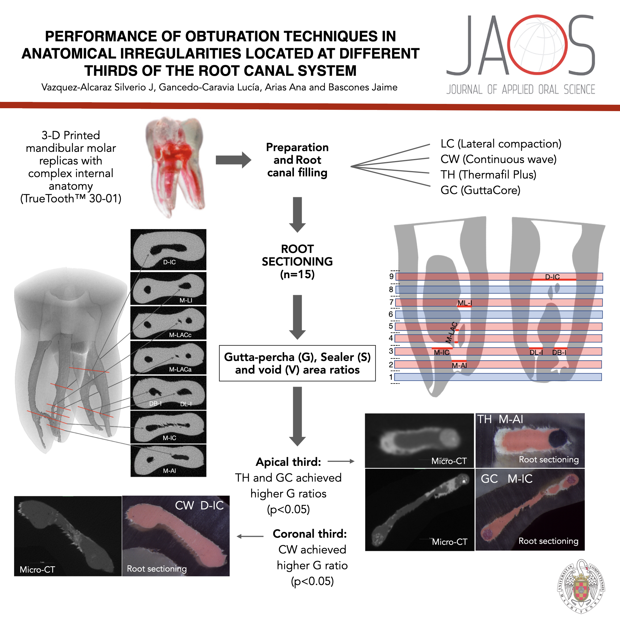
This study aimed to compare the quality of root canal obturation (ratio of area occupied by gutta-percha (G), sealer (S), and presence of voids (V)) in different anatomical irregularities (intercanal communications, lateral irregularities, and accessory canals) located at different thirds of the root canal system of mandibular molar replicas. Sixty-seven 3D printed replicas of an accessed mandibular molar were prepared using ProGlider and ProTaper Gold rotatory systems. Three specimens were randomly selected to be used as controls and did not receive further treatment. The rest were randomly distributed in 4 experimental groups to be obturated using either cold lateral compaction (LC), continuous wave of condensation (CW), and core-carrier obturation (ThermafilPlus (TH) or GuttaCore (GC)) (n=16 per group). AHPlus® sealer was used in all groups. The three controls and a specimen from each experimental group were scanned using micro-computed tomography. The rest of the replicas were sectioned at the sites of anatomical irregularities and examined at 30× magnification. The G, S, and V ratios were calculated dividing the area occupied with each element by the total root canal area and then compared among groups using the Kruskal-Wallis test. Voids were present in all obturation techniques with ratios from 0.01 to 0.15. CW obtained a significantly higher G ratio in the irregularity located in the coronal third (0.882) than LC (0.681), TH (0.773), and GC (0.801) (p<0.05). TH and GC achieved significantly higher G ratios in those located in the apical third (p<0.05). The worst quality of obturation was observed in the loop accessory canal with all obturation techniques. Whitin the limitations of this study, it can be concluded that CW and core-carrier obturation are respectively the most effective techniques for obturating anatomical irregularities located in the coronal and the apical third.
Downloads
References
Ricucci D, Siqueira JF Jr. Biofilms and apical periodontitis: study of prevalence and association with clinical and histopathologic findings. J Endod. 2010;36(8):1277-88. doi: 10.1016/j.joen.2010.04.007.
Versiani MA, Ordinola-Zapata R, Keleş A, Alcin H, Bramante CM, Pécora JD, et al. Middle mesial canals in mandibular first molars: a micro-CT study in different populations. Arch Oral Biol. 2016;61:130-7. doi: 10.1016/j.archoralbio.2015.10.020
Angerame D, De Biasi M, Brun F, Turco G, Franco V. Computed microtomography study of untreated, shaped and filled mesiobuccal canals of maxillary first molars. Aust Endod J. 2019;45(1):72-8. doi: 10.1111/aej.12286
De-deus G, Maniglia-Ferreira CM, Gurgel-Filho ED, Paciornik S, Machado AC, Coutinho-Filho T. Comparison of the percentage of gutta- percha-filled area obtained by Thermafil and system B. Aust Endod J. 2007;33(2):55-61. doi: 10.1111/j.1747-4477.2007.00047.x
Schaefer E, Schrenker C, Zupanc J, Buerklein S. Percentage of gutta-percha filled areas in canals obturated with cross-linked gutta-percha core-carrier systems, single-cone and lateral compaction technique. J Endod. 2016;42(2):294-8. doi: 10.1016/j.joen.2015.10.018
Wu MK, Ozok AR, Wesselink PR. Sealer distribution in root canals obturated by three techniques. Int Endod J. 2000;33(4):340-5. doi: 10.1046/j.1365-2591.2000.00309.x
Wolf TG, Willems L, Briseño-Marroquín B. An in vitro endodontic model to quantify the accessory canal filling potential of the vertical and lateral condensation techniques. Aust Endod J. 2021;47(2):245-51. doi: 10.1111/aej.12465
Keles A, Ahmetoglu F, Uzun I. Quality of different gutta-percha techniques when filling experimental internal resorptive cavities: a micro-computed tomography study. Aust Endod J. 2014;40(3):131-5. doi: 10.1111/aej.12043
Gok T, Capar ID, Akcay I, Keles A. Evaluation of different techniques for filling simulated c-shaped canals of 3-dimensional printed resin teeth. J Endod. 2017;43(9):1559-64. doi: 10.1016/j.joen.2017.04.029
Libonati A, Montemurro E, Nardi R, Campanella V. Percentage of gutta-percha-filled areas in canals obturated by 3 different techniques with and without the use of endodontic sealer. J Endod. 2018;44(3):506-9. doi: 10.1016/j.joen.2017.09.019
Ordinola-Zapata R, Bramante CM, Duarte MA, Cavenago BC, Jaramillo D, Versiani MA. Shaping ability of reciproc and TF Adaptive systems in severely curved canals of rapid microCT-based prototyping molar replicas. J Appl Oral Sci. 2014;22(6):509-15. doi: 10.1590/1678-775720130705.
Zhang P, Yuan K, Jin Q, Zhao F, Huang Z. Presence of voids after three obturation techniques in band-shaped isthmuses: a micro- computed tomography study. BMC Oral Health. 2021;21(1):227. doi: 10.1186/s12903-021-01584-2
Ahmed HM, Ibrahim N, Mohamad NS, Nambiar P, Muhammad RF, Yusoff M, et al. Application of a new system for classifying root and canal anatomy in studies involving micro-computed tomography and cone beam computed tomography: explanation and elaboration. Int Endod J. 2021;54(7):1056-82. doi: 10.1111/iej.13486
Dental Education Laboratories - DELabs. TrueTooth #30 [Internet]. [place unknown]: DELabs; 2020 [cited 2020 June 1]. Available from: https://delendo.com/collections/truetooth/products/truetooth-30
Holmes S, Gibson R, Butler J, Pacheco R, Askar M, Paurazas S. Volumetric evaluation of 5 root canal obturation methods in truetooth 3-dimensional-printed tooth replicas using nano-computed tomography. J Endod. 2021;47(3):485-91.e4. doi: 10.1016/j.joen.2020.11.012
De-Deus G, Reis C, Beznos D, Gruetzmacher de Abranches AM, Coutinho-Filho T, Paciornik S. Limited ability of three commonly used thermoplasticized gutta-percha techniques in filling oval-shaped canals. J Endod. 2008;34(11):1401-5. doi: 10.1016/j.joen.2008.08.015
Whitworth JM, Baco L. Coronal leakage of sealer-only backfill: an in vitro evaluation. J Endod. 2005;31(4):280-2. doi: 10.1097/01.don.0000155229.80400.fa
Gulsahi K, Cehreli ZC, Kuraner T, Dagli FT. Sealer area associated with cold lateral condensation of gutta-percha and warm coated carrier filling systems in canals prepared with various rotary NiTi systems. Int Endod J. 2007;40(4):275-81. doi: 10.1111/j.1365-2591.2006.01213.x
De-Deus G, Souza EM, Silva E, Belladonna FG, Simões-Carvalho M, Cavalcante DM, et al. A critical analysis of research methods and experimental models to study root canal fillings. Int Endod J. 2022;55 Suppl 2:384-445. doi: 10.1111/iej.13713
Reymus M, Fotiadou C, Kessler A, Heck K, Hickel R, Diegritz C. 3D printed replicas for endodontic education. Int Endod J. 2019;52(1):123-30. doi: 10.1111/iej.12964
Boutsioukis C, Arias-Moliz MT. Present status and future directions - irrigants and irrigation methods. Int Endod J. 2022;55 Suppl 3(Suppl3):588-612. doi: 10.1111/iej.13739
Tanalp J. A critical analysis of research methods and experimental models to study apical extrusion of debris and irrigants. Int Endod J. 2022;55 Suppl 1:153-77. doi: 10.1111/iej.13686
Jung M, Lommel D, Klimek J. The imaging of root canal obturation using micro-CT. Int Endod J. 2005;38(9):617-26. doi: 10.1111/j.1365-2591.2005.00990.x
Gencoglu N, Yildirim T, Garip Y, Karagenc B, Yilmaz H. Effectiveness of different gutta-percha techniques when filling experimental internal resorptive cavities. Int Endod J. 2008;41(10):836-42. doi: 10.1111/j.1365-2591.2008.01434.x
Marciano MA, Ordinola-Zapata R, Cunha TV, Duarte MA, Cavenago BC, Garcia RB, et al. Analysis of four gutta-percha techniques used to fill mesial root canals of mandibular molars. Int Endod J. 2011;44(4):321-9. doi: 10.1111/j.1365-2591.2010.01832.x
Mancino D, Kharouf N, Cabiddu M, Bukiet F, Haïkel Y. Microscopic and chemical evaluation of the filling quality of five obturation techniques in oval-shaped root canals. Clin Oral Investig. 2021;25(6):3757-65. doi: 10.1007/s00784-020-03703-9
Buchanan LS. The continuous wave of condensation obturation technique. In: Castelucci A, editor. Endodontics. Florence: Il Tridente; 2004. p. 688-701
Guess GM, Edwards KR, Yang ML, Iqbal MK, Kim S. Analysis of continuous-wave obturation using a single-cone and hybrid technique. J Endod. 2003;29(8):509-12. doi: 10.1097/00004770-200308000-00005
Liao SC, Wang HH, Hsu YH, Huang HM, Gutmann JL, Hsieh SC. The investigation of thermal behaviour and physical properties of several types of contemporary gutta-percha points. Int Endod J. 2021;54(11):2125-32. doi: 10.1111/iej.13615
Soo WK, Thong YL, Gutmann JL. A comparison of four gutta-percha filling techniques in simulated C-shaped canals. Int Endod J. 2015;48(8):736-46. doi: 10.1111/iej.12371
Iglecias EF, Freire LG, Candeiro GT, Santos M, Antoniazzi JH, Gavini G. Presence of voids after continuous wave of condensation and single-cone obturation in mandibular molars: a micro-computed tomography analysis. J Endod. 2017;43(4):638-42. doi: 10.1016/j.joen.2016.11.027
Downloads
Published
Issue
Section
License
Copyright (c) 2024 Journal of Applied Oral Science

This work is licensed under a Creative Commons Attribution 4.0 International License.
Todo o conteúdo do periódico, exceto onde está identificado, está licenciado sob uma Licença Creative Commons do tipo atribuição CC-BY.
