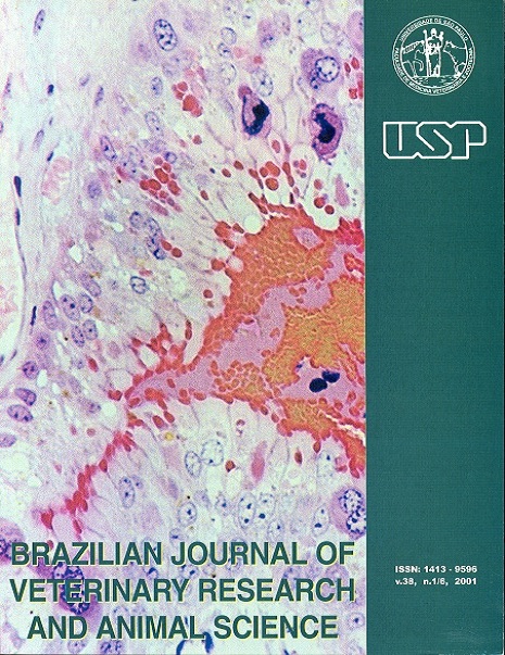
Cutaneous papillomas of green turtles: a morphological, ultra-structural and immunohistochemical study in Brazilian specimens
DOI:
https://doi.org/10.1590/S1413-95962001000200001Keywords:
Green turtles, Fibropapillomas, PapillomatosisAbstract
Eleven juvenile green turtles (Chelonia mydas) from Atlantic Ocean, Brazil, with multiple cutaneous papillomatosis were examined. Histologically, the papillomas exhibit stromal hyperplasia proliferation and epithelial proliferation. The epithelial cells had nuclear changes suggestive of viral infection and severe nuclear pleomorphism. A large nuclear halo was present in the cases of epithelial proliferation; in these cells, nuclear features were frequently dyscariotic, without inclusion. All fibropapillomas examined were negative for papillomavirus group-specific antigens (BPV) and herpesvirus group-specific antigens (HSV1 / HSV2) by the peroxidase-antiperoxidase technique. Electronic microscopy investigation was negative for papillomaviruses and herpes-viruses particles.Downloads
Download data is not yet available.
Downloads
Published
2001-01-01
Issue
Section
BASIC SCIENCES
License
The journal content is authorized under the Creative Commons BY-NC-SA license (summary of the license: https://
How to Cite
1.
Matushima ER, Longatto Filho A, Di Loretto C, Kanamura CT, Sinhorini IL, Gallo B, et al. Cutaneous papillomas of green turtles: a morphological, ultra-structural and immunohistochemical study in Brazilian specimens. Braz. J. Vet. Res. Anim. Sci. [Internet]. 2001 Jan. 1 [cited 2025 Jan. 6];38(2):51-4. Available from: https://revistas.usp.br/bjvras/article/view/5901


