
Revistas
-

V!RUS
V!RUS es una revista científica de acceso abierto (ISSN 2175-974X) anual creada en 2006 y publicada por el Nomads.usp - Núcleo de Estudios de Habitares Interactivos, del Programa de Posgrado en Arquitectura y Urbanismo, Instituto de Arquitectura y Urbanismo de la Universidad de Sao Paulo, Brasil, clasificada en el sistema científico brasileño QUALIS en el nivel A3 en una escala de A1 a B5.
V!RUS pretende constituir un locus de reflexión e interlocución en torno a temas contemporáneos relacionados con la edificación, la ciudad y la sociedad, buscando estimular ideas emergentes en diversos campos del conocimiento, como Arquitectura, Estudios Urbanos, Design, Artes, Cinema, Informática, Comunicación, Ciencias Sociales, Ambientales y Políticas y Estudios Culturales, entre otros, cuyos temas dialogan con temas de investigación de Nomads.usp.
La revista sólo acepta envíos en respuesta a convocatorias temáticas, publicadas periódicamente en su sitio web, en redes sociales y por correo electrónico a asociaciones de investigación, programas de posgrado e investigadores de Brasil y del exterior.
La revista V!RUS es una publicación académica de acceso abierto, sin fines lucrativos ni comerciales, y está protegida por la licencia CC BY-NC-SA 4.0. Tanto el servicio de publicación como el acceso a los contenidos publicados tienen carácter gratuito. La revista no cobra ningún cargo por publicar un artículo, no recibe remuneración por los servicios que presta, ni remunera a sus colaboradores.
-

Ponto Urbe
A Revista Ponto Urbe (ISSN 1981-3341) é um periódico acadêmico, online, plurilíngue, anual e gratuito organizado pelo Laboratório do Núcleo de Antropologia Urbana (LAbNAU-USP) e vinculado ao Departamento de Antropologia da Faculdade de Filosofia, Letras e Ciências Humanas da Universidade de São Paulo, Brasil.
Criado em 2007, o periódico objetiva incentivar a circulação do conhecimento científico no campo da Antropologia Urbana em diálogo com os debates antropológicos mais amplos. A Ponto Urbe tem como missão editorial proporcionar um espaço de difusão da produção acadêmica inédita realizada por pesquisadores(as) de diferentes graus de formação, regiões e instituições do país e do exterior.
A Ponto Urbe dispõe das seguintes seções: Artigos, Dossiês Temáticos, Cir-kula (voltada a artigos de outras disciplinas que dialogam com a Antropologia Urbana), Traduções (de textos raros ou atuais, não disponíveis em português), Entrevistas (com pesquisadores(as) referência na Antropologia Brasileira) e Etnográficas (relatos de campo resultados de pesquisas em andamento). As submissões são recebidas e publicadas em fluxo contínuo após a revisão por pares do tipo duplo-cego.
-
Sinopse (São Paulo)
A Sinopse – Revista de Cinema foi editada pelo CINUSP "Paulo Emilio" de 1999 a 2006, órgão da Pró-Reitoria de Cultura e Extensão Universitária da Universidade de São Paulo dedicado à exibição gratuita de filmes. Localizado no Centro Cultural Camargo Guarnieri, Cidade Universitária, traz programação variada, com mostras temáticas organizadas pela sua equipe de curadoria. Além da Sinopse, editou a Coleção Cinusp, disponível no Portal de Livros da USP.
-
Geologia e Metalurgia
Publicação do Centro Moraes Rêgo, Orgão que congrega alunos ex-alunos e professores dos Cursos de Engenheiros de Minas e Metalurgistas da Escola Politécnica da Universidade de São Paulo no período de 1945 a 1985.
-
Vértices
Vértices é a revista dos Pós-Graduandos da Área de Hebraico do Programa de Pós-Graduação em Estudos Judaicos e Estudos Árabes do Departamento de Letras Orientais da Faculdade de Filosofia, Letras e Ciências Humanas da Universidade de São Paulo.
-

Ocean and Coastal Research
Ocean and Coastal Research (OCR) é uma publicação com periodicidade em fluxo contínuo, no idioma inglês, com arbitragem por pares, destinada a divulgar resultados de pesquisa original nas diversas áreas da oceanografia, pesca, conservação marinha e ciências correlatas. Este título é uma continuação do Brazilian Journal of Oceanography (BJO), periódico publicado de forma ininterrupta desde 1950.
-
ABEI Journal
A revista é destinada a acadêmicos, pesquisadores independentes e estudantes de pós-graduação especializados em estudos irlandeses.
A revista ABEI Journal é indexada pela Cambridge Scientific Abstracts (CSA), Maryland, USA and Modern Language Association (MLA), EBSCO, Directory of Open Acess Journals (DOAJ), Diretório de Políticas Editoriais das revistas científicas brasileiras (Diadorim), Latindex e Google Scholar. A revista é publicada duas vezes por ano, em junho e dezembro, pela Associação Brasileira de Estudos Irlandeses e Universidade de São Paulo, com o suporte da Faculdade de Filosofia, Letras e Ciências Humanas, Universidade de São Paulo.
-
CINEstesia
A revista estudantil CINEstesia visa uma interdisciplinaridade ainda pouco desenvolvida na academia, cujo pilar central centra-se no cinema. Nesse sentido, a revista volta-se para aqueles que possuem pouco espaço para publicar suas produções científico-acadêmicas: os graduandos. Por isso, a CINEstesia procura disseminar produções textuais, dos mais variados campos do saber, cuja ferramenta-base - o cinema - possua função universalizante para seus leitores. A sétima arte, sob o ponto de vista deste periódico, tem caráter horizontalizante e propicia que o desenvolvimento de temas diversos se dê de forma acessível e plural. Somos um braço atuante do Núcleo de Pesquisas de Relações Internacionais (NUPRI) da Universidade de São Paulo (USP).
-
Revista BBM
A Revista BBM é publicada pela Biblioteca Brasiliana Guita e José Mindlin (BBM-USP), órgão da Pr´ó-Reitoria de Cultura e Extensão Universitária da Universidade de São Paulo. A revista empreende a tarefa de divulgação do rico acervo da Biblioteca, fruto de mais de oitenta anos de garimpo de Rubens Borba de Moraes, Guita e José Mindlin. A cada edição seguindo um tema distinto, os trabalhos de pesquisa publicados dialogam com as coleções de livros, periódicos, cartas e mapas que estão preservados no espaço. Dessa forma, trata-se de um projeto que pretente iluminar a herança histórico-cultural brasileira, através deste material que compreende o intervalo de formação do Brasil, desde os anos de colonização até o séc. XX.
-
RAUSP Management Journal
RAUSP Management Journal is a quarterly publication organized by the Business Administration department of the University of Sao Paulo (Brazil).
It is a generalist, academic journal, covering all fields of management, including Entrepreneurship; Education, Strategy and Business Economics; Corporate Governance; Finance and Accounting; Environmental Management; Public Management; Technology Management; Marketing; Quality and Productivity; Human Resources and Organizations; and Information Technology.
RAUSP Management Journal is ranked among the best Brazilian journals in Business and Management (Qualis-Capes Brazil). It is dedicated to the dissemination of research and ideas that add value to the work of scholars and practitioners in the field of Management, a mission it has been fulfilling for more than 70 years. It publishes articles selected by originality, quality, and creativity.
Original manuscripts are welcome in English or in Portuguese, provided that the authors submit an English version of the text prior to publication.
RAUSP publishes four issues per annum.
Peer review
RAUSP operates through a double blind peer review model. All articles undergo an initial assessment by the journal editor. If they are considered suitable for peer review, articles will then be reviewed by a minimum of two external reviewers to assess their suitability for publication. Final responsibility for editorial decisions rests with the journal editor.
Open access
All articles published in RAUSP Management Journal are published in Open Access under a CC BY 4.0 licence. For further information on licencing, please see the author guidelines.
Publishing Services partnership
RAUSP Management Journal is published by Emerald Group Publishing on behalf of the University of Sao Paulo (USP) and it is owned by USP. RAUSP is published under a platinum OA arrangement, in that all charges for publishing an OA article in RAUSP are funded by the University of São Paulo. There is no charge to the author.
View the journal's transparency statement.
-
Manuscrítica: Revista de Crítica Genética
Manuscrítica – Revista de Crítica Genética, ISSN 2596-2477 y Qualis A4 (evaluación brasileña de revistas, 2021-2024)*, es una publicación de la Asociación de Investigadores en Crítica Genética (Associação dos Pesquisadores em Crítica Genética APCG) y del Programa de Posgrado en Letras Extranjeras y Traducción (Pós-graduação em Letras Estrangeiras e Tradução - PPG LETRA) de la Universidade de São Paulo, realizada por la Agencia de Bibliotecas y Colecciones Digitales de la Universidad de São Paulo (ABCD). Desde 1990, publica textos que dialogan con la crítica genética, disciplina que estudia los procesos de creación en diversas áreas, como la literatura, las artes visuales, el teatro y el cine, entre otras.
* Referéncia: Plataforma Sucupira
-
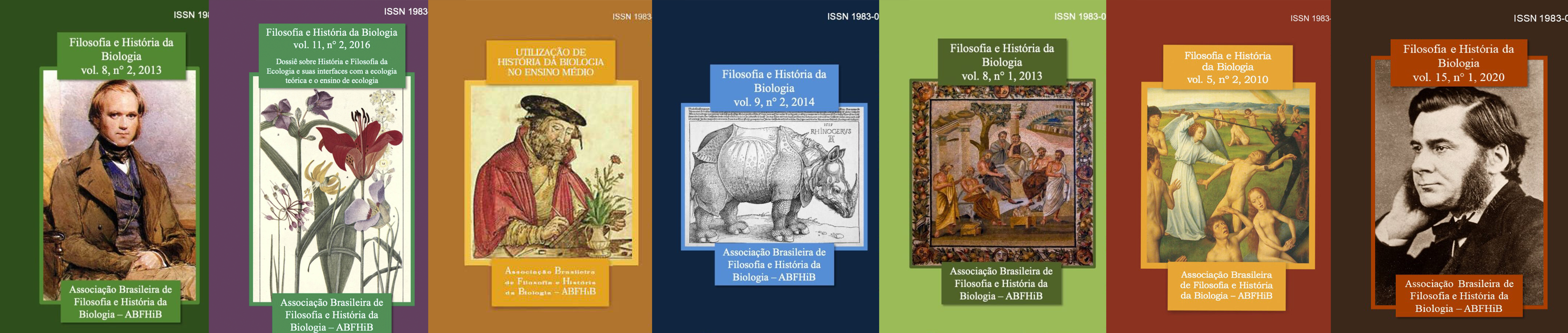
Filosofia e História da Biologia
Filosofia e História da Biologia publica artigos resultantes de pesquisas originais referentes a filosofia e/ou história da biologia e suas interfaces epistêmicas, como história e filosofia da biologia e educação científica.
-
Interfaces da Comunicação
La revista Interfaces da Comunicação está vinculada institucionalmente al Grupo de Estudos em Novas Narrativas/GENN de la Escola de Comunicações e Artes da Universidade de São Paulo, y surge de la necesidad de ampliar el campo y el área de estudios sobre las nuevas percepciones, creaciones y utilidades del concepto de narrativas. Así, la revista tiene como misión agrupar, organizar y difundir conocimientos y reflexiones sobre las narrativas en la contemporaneidad, con énfasis en los campos de la Comunicación Organizacional, las Relaciones Públicas, la Publicidad, el Periodismo, el Cine, el Turismo, la Historia, las Ciencias Sociales y las Ciencias de la Información, además de la constante interfaz con los campos de la Historia, la Psicología, la Antropología, la Sociología, la Administración, las Artes, las Literaturas, el Derecho, los Derechos Humanos, las Políticas Públicas, la Museología, la Arquitectura y el Design.
El Equipo Editorial se compone del Consejo de Redacción y del Consejo Editorial.
-
Revista de Teoria e Pesquisa Econômica
A Revista de Teoria e Pesquisa Econômica foi editada até 1970 pelo Departamento de Economia da Faculdade de Economia, Administração e Contabilidade da Universidade de São Paulo, quando passou a denominar-se Estudos Econômicos.
-
Leviathan (São Paulo)
El principal objetivo de la Revista Leviathan es ofrecer un espacio plural para la publicación de trabajos originales, reseñas, notas de investigación, traducciones y otros materiales importantes para las diferentes ramas de la Ciencia Política.
La revista está dirigida a investigadores en Ciencias Políticas, Relaciones Internacionales y campos afines, como Filosofía Política, Sociología, Economía y Estadística. En cuanto a las publicaciones, no hacemos distinciones en cuanto a metodología, enfoque o filiación institucional de los autores, y los textos se aprueban exclusivamente por su originalidad y excelencia. Salvo traducciones ocasionales, todas las obras publicadas en Leviatán son inéditas y representan contribuciones relevantes al estado del arte de la ciencia política y las relaciones internacionales.
-
Revista Ingesta
La Revista Ingesta es una publicación académica electrónica que tiene el objetivo de contribuir a la consolidación del campo de la história de la alimentación y de las drogas, a través de la divulgación de trabajos que estén dentro del debate propuesto en el área de História y disciplinas afines. De periodicidad semestral, la revista es producida por estudiantes de postgrado, miembros del Laboratorio de Estudios Históricos de las Drogas y Alimentación (LEHDA) del Departamento de Historia de la Faculdade de Filosofia, Letras e Ciências Humanas da Universidade de São Paulo (USP), Brasil.
-
Revista de Direito Mercantil
A Revista de Direito Mercantil, Industrial, Econômico e Financeiro (“RDM”) foi criada em 1951 em formato impresso e depois teve algumas edições em formato digital. Para visualização de edições anteriores pela Editora Vlex, acesse: https://livros-e-revistas.
vlex.com.br/source/revista- direito-mercantil-industrial- economico-financeiro-11164 ISSN 0102-8049
-
Zi Yue
A Zi Yue é uma revista dedicada aos estudos na área de sinologia. A submissão de trabalhos é feita em formato digital no endereço eletrônico da Revista Zi Yue, observando as regras editoriais disponíveis em em nosso site, aqui. Os artigos são submetidos a pareceristas vinculados ao domínio de conhecimento de cada trabalho.
As opiniões expressas nos artigos assinados são de inteira responsabilidade de seus autores. Todo material incluído nesta revista tem a autorização expressa dos autores ou de seus representantes legais.
-
La Junta (São Paulo)
A La Junta - Revista de Graduação em Espanhol é organizada pelas/os alunas/os de graduação e pós-graduação da área de Língua Espanhola e Literaturas Espanhola e Hispano-Americana do Departamento de Letras Modernas da Faculdade de Filosofia, Letras e Ciências Humanas da Universidade de São Paulo.
-
Revista de Graduação USP
Em versão eletrônica, a Revista de Graduação USP (Grad+) é uma publicação que passou a ser semestral em 2023, mantida pela Pró-Reitoria de Graduação da Universidade de São Paulo. O periódico, que aceita artigos inéditos, constitui-se como um espaço privilegiado de reflexão e compartilhamento de pesquisas, experiências pedagógicas e práticas de ensino que abordem o ensino e aprendizagem e temáticas afins e tragam à tona desafios e oportunidades que ocorrem ensino superior, voltadas aos cursos de graduação.
A Revista de Graduação USP tem como público alvo professores do ensino superior, pós-graduandos, professores da educação básica e profissionais da gestão educacional, bem como pesquisadores envolvidos com o tema em questão.
-
Revista da Universidade de São Paulo
Revista da Universidade de São Paulo foi publicada de forma irregular entre os anos de 1950 a 1987. A partir de 1989 passou a ser publicada como Revista USP.
-
Organicom
A cada edição, a Organicom aborda em profundidade um tema especial, organizado na forma de dossiê, trazendo reflexões sobre as tendências para a atividade e para a pesquisa em comunicação.
-

Revista da Biologia
A Revista da Biologia é um periódico científico voltado à divulgação de estudos de todas as áreas da Biologia, sem custo algum para autores e leitores. Sua criação é resultado de uma iniciativa de acadêmicos de diferentes universidades brasileiras que desejam aumentar a visibilidade dos trabalhos realizados nas diferentes áreas de Biologia, especialmente aqueles oriundos de alunos de graduação ou jovens pesquisadores.
Aceitamos submissões em inglês (preferencialmente) ou português, que contenham novidades nas áreas de Biologia, bem como revisões e métodos. Todas as publicações serão submetidas à revisão por pares. O manuscrito será publicado imediatamente após a finalização do processo editorial.
Qualis: B4
Rev. Biol., ISSN 1984-5154, DOI 10.7594/revbio
-
Mare Nostrum
Mare Nostrum es una revista sobre el Mediterráneo antiguo que desde 2010 publica el Laboratorio de Estudios en el Imperio Romano y el antiguo Mediterráneo (Universidad de San Paulo).
-
Revista ARA
La Revista ARA se dedica a reflexionar sobre Museos y Patrimonio, a partir de una discusión sobre los tiempos actuales, de acuerdo con el significado de la palabra que da nombre a la revista. Así, el pasado, el presente y lo que se espera del futuro constituyen focos privilegiados.
Para cada número, el Grupo Museo/Patrimonio (GMP) sugiere temas al Consejo Editorial (CE), formado por expertos nacionales e internacionales, quien los selecciona. También les corresponde leer las Presentaciones y respaldar a los árbitros para analizar el material enviado, observando el Reglamento.
La revista se divide en dos partes: una dedicada a las diferentes investigaciones sobre GMP en curso, denominada Dossier; el otro se centra en las Presentaciones, ambas examinadas por al menos dos expertos, con el fin de garantizar el mérito deseado.
-
Biblioteca Escolar em Revista
Biblioteca Escolar em Revista é uma revista da Faculdade de Filosofia, Ciências e Letras / USP-Ribeirão Preto que se dedica à divulgação especializada nos estudos sobre história da leitura, biblioteca escolar, práticas de leitura no âmbito escolar, literatura infanto-juvenil e mediação cultural na biblioteca escolar, publicando principalmente pesquisas originais, como também documentos especiais, traduções e resenhas.
-
Revista da Tulha
Revista académica de música y sus interfaces del Núcleo de Investigación en Ciencias de la Performance Musical (NAP-CIPEM) del Departamento de Música de la Facultad de Filosofía, Ciencias y Letras de Ribeirão Preto, Universidad de São Paulo.
-
Humanidades em diálogo
A revista Humanidades em diálogo é editada por alunos de graduação do Programa de Educação Tutorial (PET) da Universidade de São Paulo: PET Filosofia, PET História, PET Ciências Sociais, PET Sociologia Jurídica e PET Direitos. Os objetivos do periódico são contribuir para o aprofundamento do diálogo entre as diferentes disciplinas e campos do saber da área de humanidades, divulgar a produção de conhecimento elaborada por alunos de graduação, bem como proporcionar a estes alunos uma primeira experiência com publicações acadêmicas. Contamos com periodicidade anual e acolhemos trabalhos inéditos produzidos por graduandos da área de humanidades como artigos, críticas e ensaios, ilustrações e demais produções.
-

Revista de Estudos Culturais
A Revista de Estudos Culturais é uma publicação do Programa de Pós-Graduação em Estudos Culturais da Escola de Artes, Ciências e Humanidades da Universidade de São Paulo (EACH/USP). A revista incentiva a submissão de artigos originais e resenhas em todas as vertentes dos Estudos Culturais.
-
Revista Gestión & Políticas Públicas
La Revista Gestão & Políticas Públicas tiene como finalidad la publicación de artículos originales sobre temas de actualidad en la gestión de políticas públicas, preferentemente guiados por un enfoque interdisciplinario. Es una revista electrónica semestral de la Escuela de Artes, Ciencias y Humanidades de la Universidad de São Paulo (EACH-USP), en asociación con la Fundación para el Desarrollo Administrativo (FUNDAP). Los aportes a la revista pueden provenir de diferentes áreas de conocimiento, dado el amplio alcance de la gestión de políticas públicas en sus múltiples dimensiones.
La RG&PP no cobra ningún tipo de tasas o valores por el envío o la publicación de los manuscritos enviados para su evaluación y, eventualmente, publicados en nuestros volúmenes.
-
Épistémologiques
A revista Épistémologiques foi publicada entre os anos de 2000 e 2002 pelo Departamento de Filosofia da Faculdade de Filosofia, Letras e Ciências Humanas da Universidade de São Paulo.
-
Khronos
Khronos – Revista de História da Ciência é uma publicação semestral do CHC - Centro Interunidades de História da Ciência da Universidade de São Paulo (fundado em 1988) - voltada para a história e epistemologia das ciências naturais, ciências da vida, ciências humanas, técnicas e áreas correlatas. A perspectiva é interdisciplinar e visa estimular as possibilidades interpretativas dos processos de conhecimento científico e técnico em seus contextos históricos. São publicados resultados de pesquisas originais relativas a temas desde a Antiguidade até o século 21, inclusive. Além destes, são bem vindos textos de memórias de cientistas ou de instituições, bem como traduções inéditas, resenhas, notícias de projetos de pesquisa e outros assuntos de interesse para historiadores.
-
Boletim da Faculdade de Filosofia, Ciências e Letras da Universidade de São Paulo. Mineralogia
Boletim. Mineralogia - Faculdade de Filosofia, Ciências e Letras, Universidade de São Paulo
-
Opiniães
“Enfim, cada um o que quer aprova, o senhor sabe: pão ou pães, é questão de opiniães...”
(João Guimarães Rosa, Grande sertão: veredas) -
Revista Geografia Literatura e Arte
La Revista de Geografía, Literatura y Arte está vinculada al Departamento de Geografía de la Facultad de Filosofía, Letras y Ciencias Humanas (FFLCH) da Universidade de São Paulo (USP).
-

Revista Extraprensa
La revista Extraprensa es un periodico destinado a la publicación de la producción científica en las áreas de cultura y comunicación en Brasil y Latinoamérica, comprendendo temas como la diversidad cultural, ciudadanía, expresiones de las culturas populares, artes, medias alternativas, epistemología y metodología de cultura y comunicação.
Perfil de publicaciones de Extraprensa: artículos, reseñas, informe de pesquisas y crónicas científicas.
-
Boletim do Instituto de Higiene de São Paulo
O Boletim do Instituto de Higiene de São Paulo foi publicado entre anos de 1919 e 1946. O fato de ter surgido no ano seguinte à criação do Laboratório de Hygiene da Faculdade de Medicina indica a importância do Boletim como veículo de comunicação científica, legitimador e divulgador dos ideais do higienismo como matriz fundante da saúde pública no período.
Nos 28 anos em que foi editado, o Boletim apresentou características editoriais que ajudam a elucidar seu papel na legitimação da instituição à qual se vinculava e também na consolidação de uma matriz de entendimento e intervenção sobre e na saúde pública vigente. Cada um dos 88 números editados publicou um artigo, uma separata ou um comentário, e é notável sua regularidade – interrompida apenas nos anos de 1925 e 1926 –, considerando-se as potenciais dificuldades dos processos de criação, estrutura e formação institucional.
A criação do Laboratório de Hygiene em 1918, a mudança da direção do Instituto de Higiene em 1922 para Geraldo de Paula Souza, a formalização da autonomia do Instituto em relação à Faculdade de Medicina, a mudança para a sede em 1931 e a instalação da Faculdade de Saúde Pública em novembro de 1945, entre outros fatos, mostram o dinamismo da institucionalização do campo da saúde pública em São Paulo e a importância que os sujeitos históricos da época davam à comunicação científica, instrumento legitimador da identidade intelectual pretendida.
A maior parte dos trabalhos publicados são produções acadêmicas de diretores, pesquisadores, instrutores, professores e assistentes ligados ao Instituto de Higiene e também – prática comum na época – separatas publicadas em outros periódicos, trabalhos apresentados em congressos e conferências proferidas em ocasiões diversas.
Fonte: Marques, M. C. da C., & Dolci, M. de C. (2016). Boletim e Arquivos: a comunicação científica até a criação da Revista de Saúde Pública . Revista De Saúde Pública, 50, 62. https://doi.org/10.1590/S1518-8787.2016050000115
-
Intelligere
Intelligere, Revista de História Intelectual é um periódico científico semestral, eletrônico, trilingue (português, espanhol, inglês) dedicado aos estudos de História Intelectual e História das Ideias.
Intelligere publica artigos originais, entrevistas, resenhas de livros, notícias de pesquisa em andamento, traduções e fontes documentais relevantes para a história intelectual.
-
Dissenso: revista de estudantes de filosofia
A revista Dissenso foi publicada entre os anos de 1997 e 1999 pelos estudantes do curso de Filosofia, da Faculdade de Filosofia, Letras e Ciências Humanas da Universidade de São Paulo -

-
Revista LABVERDE
A Revista LABVERDE, criada em 2010 pelo Laboratório LABVERDE, com periodicidade semestral (março-agosto), tem por objetivo divulgar o andamento e resultado das pesquisas científicas, a nível de pós-graduação e promover eventos e encontros científicos, em suas áreas de atuação. Esta decisão editorial de produção somente em suporte digital teve a intenção de tornar a Revista mais ágil, facilitando tanto a colaboração quanto a leitura de pesquisadores, profissionais e demais interessados em temáticas instigantes e abordagens inovadoras na área de Arquitetura Urbanismo e Design.
-
Revista Entrecaminos
La Revista Entrecaminos es un periódico electrónico fundado en 2013 por discentes y egresados del Programa de “Pós-Graduação em Língua Espanhola e Literaturas Espanhola e Hispano-Americana” del Departamento de Letras Modernas de la Faculdade de Filosofia, Letras e Ciências Humanas (FFLCH) de la USP.
-

GIS - Gesto, Imagem e Som - Revista de Antropologia
A GIS – Gesto, Imagem e Som – Revista de Antropologia é uma revista acadêmica que engloba os campos da antropologia visual, da música e do som, da performance, do teatro e da arte.
Com vistas a criar um espaço de interlocução internacional dos materiais e reflexões produzidos por esses campos, aceitamos publicações em português, espanhol, inglês, italiano e francês, sendo que, no caso dos artigos publicados em português e espanhol, o autor deverá também providenciar a tradução do artigo para o inglês.
O bilinguismo nos artigos em espanhol e português tem por objetivo divulgar mais amplamente a produção latino-americana e de língua portuguesa.
-
Língua e Literatura
A revista Língua e Literatura está vinculada à Faculdade de Filosofia, Letras e Ciências Humanas (FFLCH) da Universidade de São Paulo (USP).
-
Estudos Japoneses
A revista Estudos Japoneses tem como missão publicar artigos de perfil acadêmico que tratem de temas relativos à Língua, Literatura e Cultura Japonesa, abordados à luz de metodologias científicas. A revista publica artigos em português, inglês, francês, espanhol e japonês.
-
Revista Música
Fundada en 1990, la REVISTA MÚSICA, ISSN 2238-7625 (Online) es una publicación semestral del Programa de Posgrado en Música de la Escuela de Comunicaciones y Artes de la Universidad de Sao Paulo (ECA/USP). La Revista publica principalmente artículos originales resultantes de investigación científica, incluyendo también otros tipos de contribuciones significativas para el área (traducciones, entrevistas, reseñas). Las contribuciones deben ser del área de investigación en Música, contemplando también sus diversas interfaces. Las reseñas deben ser de libros publicados hace menos de dos años. Las traducciones deben ser, preferencialmente, de textos clásicos del área, con innegable interés académico. Serán analizados artículos y otros trabajos inéditos en portugués, español, inglés y francés. Los autores deben consultar las normas en: https://www.revistas.usp.br/revistamusica/about/submissions
Las contribuciones pueden ser encaminados continuamente a través del Open Journal System (OJS).
Para mayores informaciones escriba a:
revistappgmus@usp.br
-
Acta Fisiátrica
A Acta Fisiátrica (ISSN 0104-7795 | e-ISSN 2317-0190) é uma publicação do Instituto de Medicina Física e Reabilitação do Hospital das Clínicas e do Departamento de Medicina Legal, Bioética, Medicina do Trabalho e Medicina Física e Reabilitação da Faculdade de Medicina da Universidade de São Paulo com o apoio da Fundação Faculdade de Medicina e da Associação Brasileira de Medicina Física e Reabilitação (ABMFR).
A Acta Fisiátrica é um periódico científico com periodicidade trimestral, de acesso livre, no formato eletrônico. Sua principal missão é difundir o conhecimento da comunidade brasileira envolvida em Medicina Física e Reabilitação, dando sempre preferência para os artigos produzidos no Brasil, porém autores de outros países também podem encaminhar sua produção científica, pois é do entendimento da revista que as contribuições estrangeiras podem fornecer novas abordagens aos problemas enfrentados no país.
-
Rapsódia
Rapsódia, um almanaque de filosofia e arte que procura mostrar as diferentes manifestações do que se costuma chamar estética.
Nos seus fascículos, ao lado de artigos sobre cinema, poética, literatura, artes plásticas, música, arquitetura e mídia eletrônica - com especial atenção à análise de fenômenos artísticos nacionais - encontram-se entrevistas, traduções, ensaios fotográficos, ficção, poesia, gravuras.
Tendo sido concebida e realizada por pós-graduandos em Filosofia da Universidade de São Paulo, rapsódia procura veicular e reunir pesquisas, contribuindo assim para uma visão de conjunto da nossa produção acadêmica em matéria de filosofia e arte. Mas a invenção artística irrompe de todos os lados, e talvez a maioria não brote em solo universitário.
-

9ª Arte (São Paulo)
9a Arte es una publicación semestral del Observatorio del Cómic de la Facultad de Comunicaciones y Artes de la Universidad de São Paulo.
-
Revista Digital de Direito Administrativo
A Revista Digital de Direito Administrativo da USP – RDDA (ISSN: 2319-0558), periódico digital e gratuito, pretende fomentar a publicação de textos de direito administrativo geral, setorial ou processo administrativo que evidenciem, de modo implícito ou explícito, a relação entre Direito, Administração Pública e o processo de desenvolvimento principalmente à luz de uma das seguintes questões centrais: como as deficiências do tratamento jurídico da Administração Pública em geral (em termos organizacionais, procedimentais, contratuais, por exemplo) ou em campos específicos (ambiente, cidades, energia, concorrência etc.) geram impactos negativos para o Estado e a sociedade? Ou, em sentido oposto, como novos institutos e reformas do direito administrativo contribuem para o bom funcionamento da Administração Pública e, em última instância, melhoram as condições de vida da sociedade?
Por conta dessa linha editorial, a RDDA estimula a submissão de artigos que tratem de novos institutos, entidades ou diplomas legais, projetos de lei em andamento, transformações do direito administrativo, deficiências do direito administrativo. Objetiva, ademais, fomentar a publicação de artigos sobre os avanços do direito administrativo estrangeiro, com ou sem análises comparativas, e preferencialmente elaborados em inglês, espanhol, italiano ou francês.
Além de abranger artigos científicos, a RDDA publica resenhas de livro, comentários sobre novidades legislativas e jurisprudenciais. Informações detalhadas sobre submissão podem ser obtidas na aba superior "SUBMISSÃO DE ARTIGOS". Artigos elaborados por alunos de graduação serão aceitos apenas excepcionalmente e desde que em coautoria com o orientador, além de relacionados, de modo preferencial, a conclusões de pesquisa financiada e aderente à linha editorial da Revista.
-

Revista de Estudios Brasileños
La Revista de Estudios Brasileños (REB) es editada por Ediciones Universidades de Salamanca en colaboración con la Universidade de São Paulo. El objetivo de la revista es fomentar y establecer discusiones académico-científicas y divulgar la producción científica sobre Brasil en las áreas de las Ciencias sociales, humanidades y jurídicas, constituyendo una plataforma en los estudios brasileños.
-
Magma
A Magma Revista é uma publicação do Departamento de Teoria Literária e Literatura Comparada da Faculdade de Filosofia, Letras e Ciências Humanas da Universidade de São Paulo. Atualmente, a revista, que inicialmente foi criada com o objetivo de divulgar a produção do corpo discente do departamento, aceita trabalhos de pós-graduandos em atividade, e pesquisadores com titulação de até cinco anos para as seções de artigos. Para as seções de criação, tradução e resenha recebemos trabalhos de graduandos em atividade de pesquisa, pós-graduandos e pesquisadores titulados, sem restrições.
-
Cadernos de Filosofia Alemã: Crítica e Modernidade
Cadernos de Filosofia Alemã: Crítica e Modernidade
Universidade de São Paulo, Brasil
ISSN Impresso: 1413-7860
ISSN Online: 2318-9800
Acceso abierto, revisión ciega
La revista Cadernos de Filosofia Alemã: Crítica e Modernidade, editada por la Universidade de São Paulo, publica en acceso abierto desde 1996 artículos, reseñas, traducciones y entrevistas de autores nacionales e internacionales. Nuestro ámbito temático abarca no sólo la filosofía alemana sino, de forma más general, las reflexiones sobre la modernidad entendida a partir del marco teórico proporcionado por la herencia de la filosofía crítica.
Esta revista fue creada por el grupo FiCeM - Filosofía Crítica y Modernidad, un grupo de investigadores de diversas universidades brasileñas, y nuestro Consejo Editorial reúne investigadores de universidades de América Latina, Estados Unidos y Europa.
Actualmente publicamos artículos, reseñas y entrevistas en portugués, español e inglés -y traducciones al portugués- en volúmenes que salen al menos semestralmente y en Dossiers especiales.
Los textos pueden enviarse en cualquier momento a través de nuestro sitio web (si tiene alguna duda, póngase en contacto con nosotros enviando un mensaje a filosofiaalema@usp.br).
-
Revista da Faculdade de Direito, Universidade de São Paulo
A Revista da Faculdade de Direito de São Paulo foi publicada entre os anos de 1893 e 1934. Com a criação da Universidade de São Paulo a Faculdade foi incorporada à USP juntamente com sua revista. A partir de então, passou a ser publicada com o título Revista da Faculdade de Direito da Universidade de São Paulo. -
Anagrama
Revista científica interdisciplinar e interinstitucional de graduação publicada pelo MIDIATO - Grupo de Estudos de Linguagem: Práticas Midiáticas do Departamento de Jornalismo e Editoração da Escola de Comunicações e Artes da Universidade de São Paulo - ECA/USP
-
Cadernos Wittgenstein
Os Cadernos Wittgenstein foram publicados entre os anos de 2000 e 2001 pelo Departamento de Filosofia, da Faculdade de Filosofia, Letras e Ciências e Humanas da Universidade de São Paulo. Em 2016 todos os números publicados foram digitalizados e disponibilizados no Portal de Revistas da USP, pelo Sistema Integrado de Bibliotecas da USP. -
Malala, Revista Internacional de Estudos sobre o Oriente Médio e Mundo Muçulmano
La revista electrónica Malala, Revista Internacional de Estudos sobre o Oriente Médio e Mundo Muçulmano, es una publicación plural abierta a cualquier persona con un trabajo original directamente sobre el Islam y el mundo musulmán o en diálogo con ellos. La revista es una iniciativa del GTOMMM (Grupo de Trabajo sobre Oriente Medio y el Mundo Musulmán) dentro del LEA (Laboratorio de Estudios Asiáticos) de la Facultad de Historia de la Universidad de São Paulo (DH/FFLCH-USP).
El proyecto de publicación nació y permanece estructurado en torno a algunas ideas la búsqueda de la interdisciplinariedad en el campo de los estudios sobre Oriente Medio y el Mundo Musulmán; la búsqueda de la definición y afirmación de un campo de estudios en la academia brasileña (sin renunciar a la inserción internacional del debate sobre el Islam , Oriente Medio y el Mundo Musulmán), y un formato innovador, plural y accesible al público más amplio, contribuyendo así a la divulgación científica y ampliando el debate, buscando un mayor diálogo con sectores de la sociedad civil, el 3er sector y actores que a menudo están involucrados con el tema, pero fuera de la academia.
En este espacio, buscamos análisis y debates relacionados con la evolución de Oriente Medio en su concepto más general (que puede incluir también el Norte de África). En cuanto al mundo musulmán, también buscamos una compresión más amplia del significado, que incluya no sólo a las sociedades musulmanas mayoritarias de Asia y África, sino también a sus minorías no musulmanas -así como a las minorías musulmanas de Europa, América y otros lugares- y su interacción con Occidente. También buscamos contribuciones sobre la propia religión, la lengua y las expresiones culturales y/o sobre cuestiones teóricas que entren dentro de nuestra órbita.
Malala, Revista Internacional de Estudos sobre o Oriente Médio e Mundo Muçulmano es una publicación que recibe contribuciones de forma continua en portugués, inglés y español. Se publican convocatorias de artículos dos veces al año, a veces con propuestas temáticas y de dossier. Los textos deben enviarse en formato Word a través del sitio web de la revista: https://www.revistas.usp.br/malala
-
Ciência e Filosofia
A revista Ciência e Filosofia foi publicada entre os anos de 1979 e 2008 pelo Departamento de Filosofia da Faculdade de Filosofia, Letras e Ciências Humanas da Universidade de São Paulo. Em 2016 todos os números publicados foram digitalizados e disponibilizados no Portal de Revistas da USP, pelo Sistema Integrado de Bibliotecas da USP. -
Autopsy and Case Reports
Autopsy and Case Reports es una revista electrónica editada por el Hospital Universitario de la Universidad de São Paulo (HU USP).Tiene como objetivo publicar artículos científicos originales, estudios clínicos o experimentales y relatos de casos que contribuyan para el desarrollo de las habilidades de raciocinio clínico, métodos diagnósticos, manejo, clasificaciones y tratamiento de enfermedades. Los artículos deberán ser preferencialmente relacionados a autopsias académicas y/o correlación clínico- patológica-radiológica, con excelente documentación de imágenes. La publicación busca difundir la información y el conocimiento científico generados por los profesionales del HU USP y de la comunidad externa e internacional de salud.Son aceptados artículos en portugués o inglés, en sistema de evaluación por los pares (double blind review). Tiene periodicidad trimestral y está siendo preparada para ser insertada en Bases de Datos de indexación bibliográfica. Dispone acceso libre a su contenido, con la finalidad de proporcionar mayor intercambio global del conocimiento científico. Se utiliza del Sistema Electrónico de Edición de Revistas, sistema de código abierto personalizado a partir del Open Journal Systems (OJS) producido por Public Knowledge Project (PKP) de la University of British Columbia, Canadá (http://pkp.sfu.ca), para construcción y gestión de publicación periódica electrónica. En Brasil es dirigido por IBICT (Instituto Brasilero de Información en Ciencia y Tecnología (http://seer.ibict.br/), del Ministerio de Ciencia y Tecnología.
-
Boletim de Botânica da Universidade de São Paulo
El boletin publica resultados de investigaciones científicas originales en cualquier campo e la Botánica, realizadas por investigadores brasileiros o extranjeros. -
Caracol
Publicación semestral del Área de Lengua Española y Literaturas Española e Hispanoamericana del Departamento de Letras Modernas de la Facultad de Filosofía, Letras y Ciencias Humanas de la Universidad de São Paulo. Único periódico de Brasil que se dedica a los estudios hispánicos. Qualis B1.
Tiene como objetivo publicar colaboraciones inéditas en español o portugués, reseñas y eventualmente textos raros que sean de interés para el debate académico dentro de las cuatro disciplinas del Área: Literatura Española, Literatura Hispanoamericana, Lengua Española y Traducción.
-
Caligrama (São Paulo. Online)
A revista Caligrama foi publicada entre os anos de 2005 a 2008 pela Escola de Comunicações e Artes da Universidade de São Paulo -

Via Atlântica
A Revista Via Atlântica, publicação do Programa de Pós-Graduação de Estudos Comparados de Literaturas de Língua Portuguesa da Universidade de São Paulo, tem por objetivo levar aos estudiosos, do Brasil e do Exterior, resultados de investigações desenvolvidas por especialistas nas áreas de Estudos Comparados de Literaturas de Língua Portuguesa, Literatura Comparada, Literatura Infantil e Juvenil, Literaturas Africanas de Língua Portuguesa, Literatura Brasileira, Literatura Portuguesa e de outras literaturas e culturas que se expressam em português. Faz parte ainda do escopo da Via Atlântica a publicação de artigos que tratem das relações interdisciplinares da Literatura com outras Linguagens e com outras Formas do Saber. A publicação abrange, além de um Dossiê temático, outros trabalhos inéditos sob a forma de Ensaios, Artigos, Entrevistas e Resenhas de livros de interesse para os Estudos Comparados de Literaturas de Língua Portuguesa e áreas correlatas. A revista Via Atlântica está inserida na área temática de Outras Literaturas Vernáculas, conforme tabela de áreas do conhecimento do CNPq (8.02.07.00-6).
-
Boletim IG-USP. Série Didática
O Boletim IG-USP. Série Didática dedicou-se a divulgar pesquisas científicas nas diversas áreas da Geologia, com publicação de artigos científicos originais, criteriosamente avaliados, sobre temática acadêmica e aplicada.
-
Brazilian Journal of Oceanography
Publicar trabajos y notas originales relacionados con los siguientes campos de la Oceanografía: oceanografía biológica, oceanografía física, oceanografía química, oceanografía geológica y pesca.
-
Cadernos de Ética e Filosofia Política
A revista Cadernos de Ética e Filosofia Política, cujo número inaugural foi lançado em 1999, tem, ao longo de mais de duas décadas, ininterruptamente e periodicamente publicado artigos dedicados à área de Filosofia. Os Cadernos visam suprir em alguma medida a demanda por textos especializados e que atendam o estado atual da questão para o campo ao qual se destina, fornecendo bibliografia a um público interessado no caráter multifacetado da reflexão sobre a ética e a política. No quadriênio 2021-2024 da Qualis Capes, a revista foi classifica no estrato A2 na área de Filosofia.
As questões relativas ao direito, à história, à religião e às artes não raro são por elas incorporadas, convertendo a um só tempo em sua matéria de investigação e seu cenário de intervenção. É esse caráter abrangente da ética e da filosofia política que lhes concede a virtude da vivacidade. Os Cadernos sempre procuraram corresponder e promover essa virtude, veiculando sobretudo a produção teórica discente, sem distinguir correntes ideológicas, linhas filosóficas ou áreas de saber incluídas nas mais diversas manifestações de reflexão.
Aqui se encontrarão artigos, ensaios, resumos de teses e dissertações, resenhas, traduções de trechos de obras e de pequenas obras. Todos os trabalhos, de recepção dos artigos, envio para pareceristas, revisão, editoração e publicação são realizados pela equipe editorial, que se reúne regularmente e toma suas decisões editoriais de modo autônomo.
A revista é editada em meio eletrônico, pelo sistema OJS, o que resulta em um ganho substancial de qualidade, pois facilita o acesso e a difusão dos textos. Somando-se a isso, contamos com um vasto corpo de pareceristas especializados nos temas, correntes filosóficas e autores enfocados pelos artigos, o que torna mais democrática a escolha dos textos destinados à publicação.
Além disso, os Cadernos utilizam o sistema de avaliação na modalidade double-blind review, nos quais todos os manuscritos enviados passam por ao menos dois avaliadores. De 2017 até o presente momento, a revista somente recebe artigos no prazo aberto de chamadas, noticiadas nesse site e em outros meios. Finalmente, desde 2015 os Cadernos têm promovido eventos na área, buscando contribuir para o debate sobre a pesquisa filosófico-política no Brasil, bem como tem editado dossiês temáticos, conduzidos, conjuntamente com o corpo editorial da revista, com editores e editoras convidados.
Convidamos os/as estudantes de pós-graduação em filosofia e pesquisadores/as interessados/as em publicar seus trabalhos a colaborar conosco, ajudando-nos a diminuir assim a distância entre a pesquisa individual e o diálogo aberto com autores e críticos.
-
Revista Alterjor
A revista Alterjor é uma publicação semestral do Grupo de Pesquisas Alterjor (ECA/USP), que tem como foco o jornalismo popular e alternativo. O primeiro termo se define pelas práticas jornalísticas realizadas em organizações do movimento social e popular, incluindo o chamado Terceiro Setor, que tenham, como objetivos centrais, o fortalecimento institucional de tais organizações e a socialização de temáticas que envolvam a defesa da cidadania e que defendam o protagonismo de segmentos sociais não hegemônicos. Já o jornalismo alternativo se conduz pelas experiências de jornalismo nas diversas mídias que tenham, como objetivo central, fomentar o debate público sobre as mesmas temáticas delimitadas na definição de jornalismo popular. Com isso, Alterjor convida o leitor a pensar sobre a viabilidade da democratização da comunicação para todos os segmentos da sociedade.
-
Revista da Faculdade de Medicina Veterinária e Zootecnia
- Continuación de la Revista da Faculdade de Medicina Veterinária, Universidade de São Paulo (1938-1971).
- Continúa como Brazilian Journal of Veterinary Research and Animal Science (1990-atual)
-
Significação: Revista de Cultura Audiovisual
Significação – Revista de Cultura Audiovisual publica artigos dedicados ao estudo dos meios e processos audiovisuais e dos sistemas digitais, pensando-os em sua diversidade de práticas e de ideias que envolvem os seus processos específicos de reflexão, criação, produção e difusão.
-
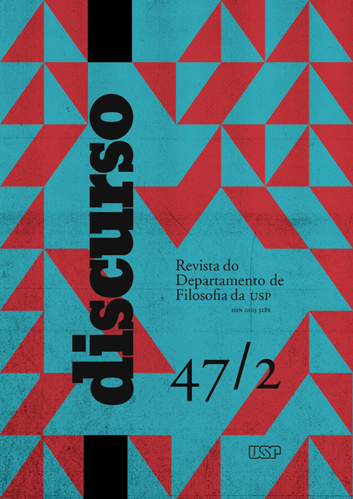
Discurso
A revista Discurso, órgão oficial do Departamento de Filosofia da USP, surgiu em 1970. Quando atravessava a mais difícil fase de sua história, atingido duramente pela violência da ditadura, este Departamento extraiu da ameaça de seu desaparecimento a força e a coragem para criar um espaço de expressão.
A revista propôs-se a veicular não apenas a produção teórica de seu corpo docente, mas também as mais diversas manifestações de reflexão sobre a cultura, sem distinguir correntes ideológicas, linhas filosóficas ou áreas do saber. Assim se pretendia garantir o pluralismo e a liberdade, numa época de obscurantismo.
ATENÇÃO
As submissões à revista Discurso encontram-se suspensas para devido tratamento dos próximos números. Pedimos aos nossos usuários e autores que acompanhem nosso quadro de notícias a fim de tomarem conhecimento sobre futuras datas de submissão.
Att.,
Equipe Editorial
-
Arquivos da Faculdade de Higiene e Saúde Pública da Universidade de São Paulo
Os Arquivos da Faculdade de Higiene e Saúde Pública da Universidade de São Paulo foram publicados entre os anos de 1947 e 1966. -
Letras Clássicas

Letras Clássicas de http://www.revistas.usp.br/letrasclassicas/index está licenciado com uma Licença Creative Commons - Atribuição-CompartilhaIgual 4.0 Internacional. -
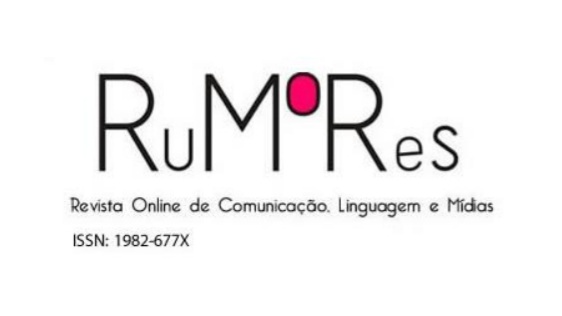
Rumores
RuMoRes – Revista Online de Comunicação, Linguagem e Mídias é um periódico científico semestral da Escola de Comunicações e Artes da Universidade de São Paulo (ECA-USP) publicado por MidiAto – Grupo de Estudos de Linguagem e Práticas Midiáticas e por Metacrítica – Rede de Pesquisa em Cultura Midiática. A revista é voltada para a divulgação de artigos científicos, resenhas e entrevistas que contribuam para o debate sobre crítica de mídia e comunicação, com textos originais e inéditos (de autoria individual ou coletiva) de autores/as com titulação mínima de doutor/a ou doutorando/a, vinculados a instituições de ensino superior, recebendo-os em sistema de fluxo contínuo.
Os artigos devem ser encaminhados em Word, fonte TNR 12, espaçamento 1,5, seguindo as orientações encontradas nas Normas de Publicação. A revista conta com apoio da Agência de Bibliotecas e Coleções Digitais da Universidade de São Paulo e do Programa de Pós-Graduação em Meios e Processos Audiovisuais da ECA-USP.
Contato: rumores@usp.br
Informações: midiato@usp.br
MidiAto: www.usp.br/midiato
Rede Metacrítica: https://midiato.wordpress.com/metacritica-rede-de-pesquisa-em-cultura-midiatica/
-
Estudos Semióticos
Publicação quadrimestral online do Programa de Pós-Graduação em Semiótica e Linguística Geral da FFLCH-USP, a revista Estudos Semióticos (ISSN 1980-4016) veicula, em acesso aberto, trabalhos da área de Semiótica, bem como dos campos limítrofes, dirigidos à comunidade dos pesquisadores. Podem ser propostos trabalhos que lidem com os signos, os textos, os discursos e as práticas sociais produtoras de sentido, desde que sejam inéditos e dialoguem com as teorias semióticas. Admitem-se trabalhos em português, francês, inglês, espanhol e italiano.
Em cada temporada anual, a revista publica três números: um de tema livre e dois com dossiês temáticos. Eventualmente, pode ser publicada também uma edição extra no formato de um dossiê especial.
No último evento de classificação do Qualis Periódicos da CAPES (2024), a revista foi classificada no estrato A2.
-
Clinical and Laboratorial Research in Dentistry
A Clinical and Laboratorial Research in Dentistry é uma publicação com sistema de fluxo contínuo, com revisão por pares, com o objetivo de divulgar novos conhecimentos de interesse para pesquisadores e clínicos em Odontologia e áreas relacionadas. A avaliação dos textos apresentados levará em consideração a originalidade, relevância e a metodologia utilizada. A revista é dedicada à difusão de conhecimentos e informações relevantes para Odontologia. A CLRD é gratuita desde a submissão até a publicação. Nós encorajamos fortemente as submissões em inglês.
A revista está indexada nas seguintes bases de dados:


Curta nossa página no Facebook e acompanhe as novidades!
https://www.facebook.com/clresearchindentistry
-
InCID: Revista de Ciência da Informação e Documentação
InCID: Revista de Ciência da Informação e Documentação es una publicación de acceso abierto, publicada por la USP-Ribeirão, que se dedica a la divulgación especializada en el área de la información, publicando principalmente investigaciones originales, así como documentos especiales, traducciones y reseñas.
-
Journal of Information Systems and Technology Management
La misión de JISTEM es publicar pesquisas relevantes para la gestión de la tecnologia y sistemas de información en las organizaciones y en la sociedad.
-
Agrária (São Paulo. Online)
A revista Agrária tem como proposta a construção de um canal de debate em torno da questão agrária, em uma perspectiva crítica, com o compromisso de contribuir para a compreensão de seus fundamentos e suas diferentes formas de manifestações territoriais.
Nesse sentido a revista debate temas atuais apresentados geralmente sob a forma de dossiês, com contribuições de pesquisadores nacionais e internacionais. A revista possui as seguintes seções: Artigos, Teoria em Debate e Resenhas.
-
MATRIZes
MATRIZes es una revista para la publicación de producción científica cuyo objeto de estudio es la comunicación. Alberga trabajos teóricos, experiencias de análisis y formulaciones conceptuales sobre procesos comunicativos, medios, mediaciones y emergencias de interacciones en la sociedad de la información generalizada contemporánea. Es una revista abierta a reflexiones sobre las transformaciones históricas de las mediaciones en la cultura; sobre la producción de lenguajes y sus interfaces; sobre las implicaciones sociopolíticas de las actividades implementadas, así como sus consecuencias cognitivas. Con este fin, conserva el horizonte inter y transdisciplinario de las contribuciones teóricas y metodológicas del pensamiento comunicacional. Por lo tanto, se espera cambiar el tamaño del conocimiento histórico y las tradiciones que contribuyen a definir, mapear y explorar conceptualmente eventos comunicativos, reviviendo los compromisos que también son parte de la historia de PPGCOM-ECA-USP. En el límite, es evidente la necesidad de crear un espacio para la construcción de una teoría crítica y consecuente de las prácticas de estudio de la comunicación.
-
Anais do Museu Paulista: História e Cultura Material
Publicar artículos teóricos y monográficos que se basen en prácticas sociales mediadas por la materialidad y que se aborden como cuestiones históricas, museológicas y de conservación.
-
Brazilian Journal of Latin American Studies
A Brazilian Journal of Latin American Studies (Cadernos Prolam/USP) é uma revista científica especializada em difundir conclusões de pesquisa, análises e interpretações, bem como pensamento e teorias sobre a América Latina. Criada em 2002, pelo Programa de Pós-graduação Integração da América Latina (PROLAM/USP), desde sua criação é uma revista voltada para autores e público de nível de pós-graduação. Na fase inicial o foco das publicações da BJLAS era as relações internacionais. Com o passar dos anos, o periódico ampliou seu universo disciplinar e temático, e hoje publica trabalhos nos diversos campos das humanidades, artes e ciências sociais.
Assim, tem como eixo organizador incluir temáticas (a) de impacto regional para a América Latina ou (b) trabalhos com metodologias comparativas sobre dois ou mais países deste continente.
Considera-se que os manuscritos devem contribuir de modo significativo ao avanço do conhecimento científico em temáticas sensíveis à América Latina, por este motivo, as propostas publicadas são elaboradas por autores com nível de pós-graduação. As problemáticas que tratam de América Latina exigem perspectivas transdisciplinares com abordagens sobre tópicos transversais em questões sociais, políticas, econômicas, jurídicas, históricas, culturais, artísticas, de comunicação social.
Finalmente, a BJLAS tem interesse em divulgar resenhas de livros recentemente publicados ou de obras de grande relevância para a região, como clássicos do pensamento latino-americano, e aceita também críticas de arte ou ensaios de qualidade.
-
Arquivos de Zoologia
- Arquivos de Zoologia, continuación de “Arquivos de Zoologia do Estado de São Paulo”, v.1 (1940) - v.14 (1966), es un periódico tradicional en el área de Zoología en Brasil.
- La revista tiene como objetivo publicar trabajos científicos originales en las áreas de sistemática, paleontología, biología evolutiva, ecología, taxonomía, anatomía, comportamiento, morfología funcional, biología molecular, ontogenia, estudios faunísticos, biogeografía e historia de museos y colecciones de historia natural.
- Publicada por el Museu de Zoologia da Universidade de São Paulo (MZUSP), la revista es indexada en las principales bases de referencia y se muestra cada 6 (seis) meses.
-
Resenhas do Instituto de Matemática e Estatística da Universidade de São Paulo
A revista Resenhas do Instituto de Matemática e Estatística da Universidade de São Paulo foi publicada entre os anos de 1993 e 2005. A partir desta data continuou a ser publicada com o título São Paulo Journal of Mathematical Sciences -
Pesquisa em Educação Ambiental
Aportar contribuciones para la consolidación y fortalecimiento del campo de la investigación en educación ambiental, por intermedio de la publicación de artículos inéditos que proporcionen subsidios para el progreso teórico y práctico de la educación ambiental.
-
Pesquisa Odontológica Brasileira
A revista Pesquisa Odontológica Brasileira foi publicada entre os anos de 2000 e 2003 e após essa data passou ser editada com o título Brazilian Oral Research.
-
Boletim IGA
Divulgar las investigaciones científicas para la publicación de artículos científicos originales, evaluados con criterio sobre la temática académica y aplicada en las diversas áreas de la Geologia. -
Psicologia USP
Publicar trabajos originales que pueden contribuir al conocimiento y desarrollo de la Psicología y ciencias afines, tal como artículos teóricos y ensayos enfatizando algunos temas clásicos como Memoria, Familia, Conciencia, Insensibilidad, y Alteridad. -
Revista Brasileira de Ciências Farmacêuticas
Divulgar os mais recentes avanços da área das Ciências Farmacêuticas, através da publicação de artigos originais, notas prévias, trabalhos de revisão e resenhas. -
Revista de Estudos Orientais
A Revista Estudos Orientais é uma publicação do Departamento de Letras Orientais da Faculdade de Filosofia, Letras e Ciências Humanas da Universidade de São Paulo. As linhas de pesquisa da Revista concentram-se nos estudos linguísticos, literários e culturais das áreas constituintes do Departamento: Árabe, Armênio, Chinês, Coreano, Hebraico, Japonês e Russo. Outras áreas ligadas aos estudos asiáticos e eslavos completam esse escopo. São aceitos para publicação artigos, resenhas, traduções e entrevistas relacionadas às áreas de pesquisa mencionadas.
A Revista de Estudos Orientais está retomando as suas atividades com uma nova equipe, e cumprindo com todas as exigências acadêmicas necessárias para uma rápida atualização e evolução do seu conceito.
-
Celeuma
Celeuma é uma palavra estranha e um pouco esquecida. Ela significa “discussão intensa”, “agitação ruidosa”. Não são brigas ou gritos, contudo, que o leitor deve esperar do material da nova revista, publicação eletrônica que terá três números por ano e que agora é lançada nas comemorações dos 20 anos do Centro Universitário Maria Antonia vinculado à Pró-Reitoria de Cultura e Extensão Universitária da Universidade de São Paulo.
O tema de Celeuma são as artes na atualidade: a crítica, as teorias da ficção e da narrativa, os impactos da tecnologia, a mistura cada vez mais complexa entre as diferentes linguagens. E aí, em seus assuntos, se confundem estranhezas e esquecimentos, discussões e ruídos. É um campo vasto de pesquisa e conversa, para alguns fundamental e constitutivo, para outros insignificante e instrumentalizável, e, até por essa disparidade de percepções, cada vez mais urgente e necessário. Daí esse nome: este o seu propósito.
-
Cadernos CERU
Divulgar investigaciones científicas de investigadores nacionales e internacionales de la área de Ciencias Humanas.
-
Revista da Faculdade de Medicina Veterinária, Universidade de São Paulo
1938-1971: A Revista da Faculdade de Medicina Veterinária é publicada sob a forma de fascículos que serão reunidos em volume e não tem data certa de aparecimento.
Título abreviado: Rev. Fac. Med. Vet.
ISSN: 0301-7273
eISSN: 2318-5066- Continua como: Revista da Faculdade de Medicina Veterinária e Zootecnia da Universidade de São Paulo (1972-1989).
ISSN: 0303-7525
e-ISSN: 2318-3659
- Continua como: Revista da Faculdade de Medicina Veterinária e Zootecnia da Universidade de São Paulo (1972-1989).
-
Caderno de Estudos
La misión del Cadernos de Estudos es la divulgación de producción científica relevante en el área de Contabilidad, Contraloría, Actuaría y Finanzas, producida por profesores, investigadores, alumnos y profesionales de Brasil y del extranjero, seleccionada exclusivamente basado en calidad y contribución efectiva al desarrollo del conocimiento en ese campo, a la luz de opiniones de evaluadores. -
Journal of Ancient Philosophy
En la Revista de Filosofía Antigua se publican artículos, reseñas y notas críticas sobre la filosofía antigua greco–romana, al igual que traducciones de textos clásicos al portugués y al español. Además, provee información acerca de congresos, encuentros y conferencias acerca de la filosofía antigua greco–romana. Es publicada dos veces por año, en Mayo y en Octubre.
La Revista de Filosofía Antigua fue creada con el fin de fomentar los estudios clásicos en América Latina, sirviendo como un medio para que los especialistas en filosofía antigua publiquen sus trabajos y discutan temas de interés común. Igualmente, con ella se pretende promover el diálogo internacional entre diferentes lenguajes y aproximaciones. Está abierta a los investigadores del mundo entero, que quieran presentar sus contribuciones para ser incluidas en esta revista. Los idiomas aceptados son: portugués, español, inglés, francés, alemán e italiano. Todas las contribuciones son leídas por evaluadores anónimos.
Indexeda en: Catálogo LatinIndex, EBSCOhost, Portal de Periódicos da CAPES, Philosopher's Index, Qualis CAPES

Esta obra está bajo una Licencia Creative Commons Atribución 4.0 Internacional. -
Teresa
Teresa – Revista de literatura brasileira (Qualis B2), uma publicação da área de Literatura Brasileira da Universidade de São Paulo (USP), criada em 2000, tem como objetivo publicar pesquisas e investigações, abordando a área de Literatura Brasileira em perspectivas originais e avançadas, desenvolvidas por pesquisadores no Brasil e no exterior.
-
Educação e Pesquisa
Educação e Pesquisa es una publicación de la Faculdade de Educação de la Universidade de São Paulo - FEUSP. A partir de 2018, se edita de forma continua en volumen único anual. Publica artículos inéditos en el área de educación, en portugués, español e inglés, resultados de investigaciones de carácter teórico o empírico, o revisiones de literatura, caracterizadas por el diálogo consistente con el campo de la educación y el área de estudios en que se inscriben, por la asignación del trabajo conceptual y metodológico, por la descripción de procesos analíticos, así como por la relevancia de las contribuciones para el desarrollo de los conocimientos en Educación.
-
Revista de Saúde Pública
Publicar y diseminar productos del trabajo científico relevantes para la Salud Pública.
-
Revista aSPAs
Revista del Programa de Pos-Graduación en Artes Escénicas - Universidad de São Paulo
-
Estudos Avançados
Su propuesta central es transformar el conocimiento y la crítica en impulsores del progreso social, no solo de los brasileños sino de todos los pueblos en vías de desarrollo.
-
Revista do Instituto de Estudos Brasileiros
El Instituto de Estudios Brasileños de la Universidad de San Pablo es un centro multidisciplinario de educación, investigación y documentación sobre la cultura del país. Reúne profesionales de una variada formación que ofrecen materias de grado, posgrado y extensión y orientan trabajos de iniciación científica, maestrías y posdoctorados. Reflexionar sobre la sociedad brasileña articulando múltiples áreas del saber constituye el desafío fundante del IEB. Esto motivó, a lo largo de los años, desde su fundación en 1962 por Sérgio Buarque de Holanda, la producción de una infinidad de investigaciones originales; parte de las cuales encontró un espacio en su Revista. Creada en 1966, la Revista del Instituto de Estudios Brasileños de la USP fue editada, sin interrupciones, hasta 1997. Por su propia naturaleza, el Instituto siempre tuvo un carácter multidisciplinario acogiendo diferentes tendencias y líneas de investigación y abriendo sus páginas a investigadores de una enorme diversidad de instituciones. En muchas áreas del conocimiento, la revista formó parte del movimiento de renovación de los estudios brasileños a partir de los años '60 y fue uno de los principales vehículos de difusión del conocimiento científico originado en las universidades brasileñas y de sus programas de posgrado entre 1970 y 1990. Por otra parte, fue uno de los medios de divulgación del vasto acervo del IEB, que hoy incluye las colecciones personales de nombres fundamentales de la cultura brasileña, tales como Mário de Andrade, Graciliano Ramos, Guimarães Rosa, Osman Lins, Alberto Lamego, Yan de Almeida Prado, José Honório Rodrigues, Caio Prado Jr., Pierre Monbeig, Anita Malfatti, Francisco Mignone e Camargo Guarnieri. La Revista del IEB, espacio de debate destinado a abarcar una multiplicidad de temas, corrientes de pensamiento y líneas de investigación, resurgió en septiembre de 2006 con su propuesta integradora de las múltiples ramas de las Ciencias Humanas, asumiendo el desafío de pensar a Brasil de una forma crítica y facilitar el diálogo entre las áreas del conocimiento, procurando conservar, sin embargo, las especificidades y el rigor técnico de lecturas especializadas. Se trata, por lo tanto, de presentar estudios cuyos resultados, por su relevancia o significado teórico y metodológico pueden o deben trascender fronteras. Además, y con el mismo propósito de acoger debates en los que se confronten saberes de diversas procedencias, la Revista del IEB abre sus páginas tanto para el análisis de los problemas del Brasil contemporáneo como para las reflexiones que investiguen las dimensiones teóricas de los estudios brasileños. En este sentido, enfrenta el desafío de convertirse en un espacio aglutinador - pero no armonizador - de las innumerables fronteras del conocimiento.
-
Revista de Italianística
A Revista de Italianística é um periódico do Programa de Pós-Graduação em Língua, Literatura e Cultura Italianas da Universidade de São Paulo. A revista publica semestralmente números dedicados aos estudos linguísticos e estudos literários para divulgar a produção científica de pesquisadores nacionais e estrangeiros ligados à área da italianística.
-
Fisioterapia e Pesquisa
Informaciones básicas
La revista Fisioterapia e Pesquisa (ISSN 1809-2950), es la continuación de la Revista de Fisioterapia de la Universidad de São Paulo (ISSN 1413-7879), es una revista científica creada en 1994, cuya misión es difundir la producción científica y el avance del conocimiento en el campo de la fisioterapia y rehabilitación y disciplinas afines. Em aspectos relacionados com la investigación básica, clínica y aplicada.
Frecuencia: trimestral
Fuentes de indexación
SCIELO- Scientific Electronic Library Online
BVS – Biblioteca Virtual em Saúde
CINAH - Cumulative Index to Nursing and Allied Health Literature
LILACS – Latin American and Caribbean Health Sciences
LATINDEX – Sistema Regional de Información em Linea para Revistas Cientificas de Américas
Sport DISCUS
Propriedad intelecutal
Todo el contenido de la revista, excepto donde está identificado, está licenciado bajo una Licencia Creative Commons del tipo atribución BY.
Patrocinadores
La publicación recibe financiación de:
AGUIA- Agência USP de Administración de la Información Acadêmica
FMUSP – Faculdad de Medicina de la Universidad de São Paulo
CREFITO – Consejo de Fisioterapia e Terapia Ocupacional
-
Revista do Museu de Arqueologia e Etnologia
La Revista do Museu de Arqueologia e Etnologia fue creada en 1991, en sustitución de las revistas del antiguo Museu de Arqueología e Etnología, Instituto de Pré-historia y Museu Paulista (Dédalo, Revista de Pré-História y Revista do Museu Paulista), como fruto de la fusión de instituciones vinculadas a la investigación en Arqueología y Etnología. Es una revista académica dedicada a la publicación de trabajos sobre Arqueología, Etnología y Museología, con énfasis en África, América, el Mediterráneo y Oriente Medio. En 2016, todos los números publicados fueron digitalizados y puestos a disposición en acceso abierto en la USP Portal de Revistas.
-

-
São Paulo Journal of Mathematical Sciences
Publicar artículos de alto nivel en todas las áreas de la matemática, estadística y ciencia de la computación en las cuales nuestros investigadores han trabajado, de forma a presentar un retrato vívido de la vida científica de nuestro instituto. -
Boletim IG-USP. Publicação Especial
O Boletim IG-USP. Publicação Especial teve como objetivo publicar assuntos temáticos diversos, abrangendo revisões que demandam trabalhos mais longos e as contribuições científicas que fazem parte de eventos.
-
Journal of Human Growth and Development
La revista Journal of Human Growth and Development (anteriormente titulada Revista Brasileira de Crescimento e Desenvolvimento Humano) es una revista de libre acceso, revisada por colegas, que publica artículos originales de investigación y artículos de revisión en diversas áreas de las ciencias de la vida.
La revista Journal of Human Growth and Development fue fundada en 1991 por el Profesor Arnaldo Augusto Franco de Siqueira. link: http://revistas.marilia.unesp.br/index.php/jhgd
-
Signos do Consumo
La Revista Signos do Consumo es especializada en los estudios de la publicidad y de la propaganda, relaciones públicas y en los fenómenos del mercado y del consumo, y tiene como misión crear un espacio de intercambio de conocimientos en red, por medio de artículos científicos y reseñas de libros, para la comunidad científica nacional e internacional, interesada en divulgar los resultados de sus investigaciones, ensayos y reflexiones referentes a los estudios del área de Comunicación. Idiomas de los artículos para la publicación: Portugués, Español e Inglés.
ISSN: 1984-5057
-
Literartes
LITERARTES é revista do grupo de estudos Produções Literárias para crianças e jovens da área de Literatura Infantil e Juvenil da FFLCH/USP. Publicação periódica - em princípio, semestral - que visa a propiciar um espaço de reflexões sobre a arte literária e os diálogos que vem estabelecendo com outras linguagens, outras artes e suportes.
-
Revista Crioula
A Revista Crioula é uma publicação científica dos alunos de pós-graduação da Área de Estudos Comparados de Literaturas de Língua Portuguesa do Departamento de Letras Clássicas e Vernáculas da Universidade de São Paulo (ECLLP - DLCV - FFLCH/USP).
O periódico promove a difusão da produção científica dos alunos e ex-alunos do Programa de Pós-Graduação em Estudos Comparados de Literaturas de Língua Portuguesa da USP, bem como de pesquisadores de outras universidades do Brasil e do exterior.
-
Non Plus
Revista da Área do Francês da FFLCH – USP
A Non Plus é uma publicação eletrônica semestral do programa de Pós-Graduação em Estudos Linguísticos, Literários e Tradutológicos em Francês, do Departamento de Letras Modernas, da Faculdade de Filosofia, Letras e Ciências Humanas(FFLCH) da Universidade de São Paulo(USP).
A Non Plus publica artigos de caráter teórico que contemplem áreas cujo objeto de estudo seja a linguagem em suas diversas manifestações, tais como os segmentos de Didática, Estudos Literários, Língua e Tradução. Além disso, traz Resenhas de teses e dissertações defendidas relativas às áreas mencionadas, no intuito de difundir a produção acadêmica dedicada às Letras francesas no Brasil.
-

Novos Olhares
Novos Olhares – Revista de Estudos Sobre Práticas de Recepção a Produtos Midiáticos, foi criada em 1998 pelo Grupo de Estudos Sobre Práticas de Recepção a Produtos Mediáticos, coordenado por Mauro Wilton de Sousa, Professor Titular da Escola de Comunicações e Artes da Universidade de São Paulo, que se tornou seu editor. Até 2007, quando sua publicação foi descontinuada, a revista contou com 20 edições impressas, tornando-se a principal publicação do país no campo dos Estudos Culturais e contando com a contribuição de renomados pesquisadores nacionais e internacionais.
A partir de 2012, a revista ressurgiu como publicação online vinculada ao MidiaSon: Grupo de Estudos e Produção em Mídia Sonora, da Escola de Comunicações e Artes da Universidade de São Paulo. Eduardo Vicente, Professor Associado da instituição, assumiu, desde então, a função de editor da publicação, que teve o seu Comitê Editorial completamente renovado. Para a retomada, optou-se pela mudança na numeração da revista, que foi reiniciada no formato volume/número. Assim, as edições de 2012 tornaram-se, respectivamente, os números 1 e 2 do Volumes 1.
Desde 2013, a revista é hospedada no Portal de Periódicos da Universidade de São Paulo no endereço www.revistas.usp.br/novosolhares. Além de todas as edições online, são disponibilizadas também as versões digitalizadas das 20 edições impressas da revista que, além de trazer contribuições de autores como Bernard Miège, Lorenzo Vilches, Jesús Martín-Barbero e Antonio Fausto Neto, entre outros, reúnem entrevistas de pesquisadores como Octávio Ianni, Renato Ortiz, Sérgio Adorno, Eduardo Peñuela-Canizal, Arlindo Machado e Maria Lourdes Motter, entre outros.
A partir de 2025, a Novos Olhares passou a ser publicada em fluxo contínuo, com uma única edição/volume anual.
A revista publica, em suas edições, contribuições voltadas a um amplo leque de temáticas dentro da área de Comunicação. Sem abandonar sua vocação inicial, ligada às praticas de recepção, o periódico tem se tornado uma referência também na publicação de trabalhos voltados a diferentes aspectos da produção audiovisual, com destaque para o rádio, o podcast e a música popular industrializada.
-
Paisagem e Ambiente
Apresentar y discutir temas ligados a los estudios de los Espacios Libres de Edificacción e del Ambiente, divulgando la produccion cientifica y professional brasileña y internacional.
-
Plural
Plural es una publicación académica electrónica bianual, coordinada y editada por investigadores del Programa de Posgrado en Sociología de la Facultad de Filosofía, Lenguas y Humanidades de la Universidad de São Paulo (FFLCH-USP), con clasificación Qualis B2 en el cuatrienio 2013-2016.
-
Pandaemonium Germanicum
La revista Pandaemonium Germanicum, publicada desde 1997 por el Área de Alemán del Departamento de Letras Modernas de la FFLCH/USP y por el Programa de Posgrado en Lengua y Literatura Alemana, constituye un foro de discusión académica en los campos de la Germanística, es decir: literaturas de lengua alemana, literatura comparada y estudios culturales vinculados a los países de lengua alemana, lingüística alemana, lingüística aplicada y contrastiva (portugués/alemán), enseñanza del alemán como lengua extranjera/adicional y estudios traductológicos.
Pandaemonium pretende contribuir a la divulgación de investigaciones de germanistas brasileños y de otros países, así como al diálogo entre la Germanística, las demás áreas de Letras y otras áreas del conocimiento.
Pandaemonium es una revista de acceso abierto, editada semestralmente hasta 2015 y tres veces al año desde 2016. Los artículos se someterán a una revisión por pares de doble ciego (double blind peer review). En caso de evaluaciones divergentes, se recurrirá a una tercera evaluación crítica; si se aprueba el artículo, el artículo aún pasará por una revisión textual detallada.
-
Acolhendo a Afabetização em Países de Língua Portuguesa
Publicar artículos inéditos, resultantes de investigación, que contengan la información pertinente sobre los países cuyo idioma oficial es el portugués, a fin de que el conocimiento sobre lo que está sucediendo en la educación en esos lugares puedan ser accesibles a la comunidad interesada en la alfabetización. -
Anais da Escola Superior de Agricultura Luiz de Queiroz
Publicar artículos originales que contribuyan para o desarrollo científico de las Ciencias Agrarias. -
ARS
La revista Ars es una publicación del Programa de Posgrado en Artes Visuales de la Escuela de Comunicaciones y Artes de la Universidad de São Paulo que reúne trabajos relevantes en el debate del arte producidos en el medio universitario y fuera de él. Propone, además, un enfoque ampliado a las exigencias de la propia producción artística contemporánea. Así pues, tiene como objetivo contribuir al debate cultural más allá de los muros de la universidad e interrogar las propias perspectivas del arte en el contexto contemporáneo. Al mismo tiempo valora el área teórica y la contribución de disciplinas más antiguas, como la filosofía, la estética y la historia del arte, aunque frecuentemente se intenten confrontar o señalar los limites de esta tradición frente a los desafíos de la situación contemporánea. Ars se interesa en divulgar, junto a los trabajos de los artistas, críticos, historiadores de arte y alumnos de posgrado del Departamento de Artes Visuales, colaboraciones de investigadores, artistas, intelectuales y otros profesionales del medio artístico, a favor de la experimentación y la investigación teórica especializada.
Ars cuenta, actualmente, con versión electrónica (ISSN 2178-0447).
La revista es indexada en SciELO, LatinIndex, DOAJ y Portal de Revistas de la USP.
Clasificación Qualis/Capes para área de Artes/Música: A1
-
África
Lançada em janeiro de 1978, a Revista África é um periódico internacional e bilíngue do Centro de Estudos Africanos da Faculdade de Filosofia, Letras e Ciências Humanas da Universidade de São Paulo (CEA FFLCH-USP).
A Revista África, de publicação anual e fluxo contínuo, contribui na divulgação de estudos sobre o continente africano. Aceita trabalhos inéditos, resenhas, ensaios, notas de leitura, artigos ou notícias relacionados à realidade africana, afro-brasileira e da diáspora negra em geral, nas especialidades de sociologia, antropologia, ciência política, relações internacionais, geopolítica, história, literatura, música e saúde.
-

Pós. Revista do Programa de Pós-Graduação em Arquitetura e Urbanismo da FAUUSP
La revista Pós fue creada como un canal de comunicación más amplia de esta comunidad científica, tanto en el ámbito nacional cuanto internacional, así como para los investigadores de las diversas áreas académicas que se relacionan con el universo de la arquitectura y de la ciudad, con la inteción de registrar la memoria del pensamiento arquitectónico, de hacer circular de manera ágil los resultados de las encuestas y de mantener el debate lo más actualizado posible.
-
Boletim do Instituto Oceanográfico
O Boletim do Instituto Oceanográfico destinou-se a publicar trabalhos originais e de real valor científico, voltados à oceanografia e tecnologia dos produtos do mar e de outras ciências correlatas.
-
Boletim IG-USP. Série Científica
Divulgar las investigaciones científicas para la publicación de artículos científicos originales, evaluados con criterio sobre la temática académica y aplicada en las diversas áreas de la Geologia. -

GEOUSP Espaço e Tempo (Online)
Revista del Programa de Posgrado en Geografía Humana y el Programa de Posgrado en Geografía Física
Departamento de geografía
Facultad de Filosofía, Letras y Ciencias Humanas.
Universidad de San Pablo
Brasil
ISSN: 2179-0892 -
Brazilian Journal of Veterinary Research and Animal Science
Publicar artículos completos, notas previas y artículos de revisión en las áreas de medicina veterinaria, zootecnia y ciencias afines, elaborados por especialistas nacionales o extranjeros.
-
Cadernos de Psicologia Social do Trabalho
Cadernos de Psicologia Social do Trabalho (CPST) - Cuadernos de Psicología Social del Trabajo (CPST) - es una revista de publicación semestral, online, de acceso abierto y de carácter interdisciplinario. El periódico tiene como objetivo difundir la producción científica en el campo del trabajo y de los procesos organizativos, a partir de las lecturas de la psicología social y de otras disciplinas de las ciencias humanas y sociales, que se sitúen en el ámbito de las perspectivas críticas. Siguiendo esa línea, se destaca la centralidad de la categoría trabajo y la importancia de favorecer el punto de vista de los trabajadores, a diferencia de enfoques gerenciales, que los consideran solo como recursos. La revista prioriza la publicación de artículos que presenten reflexiones sobre situaciones concretas y que contribuyan con procesos de transformación, dirigidos a la promoción de la salud y a la garantía de derechos. Son bienvenidas las contribuciones apoyadas en diferentes lecturas teóricas. CPST publica artículos originales como relatos de investigaciones, intervenciones, experiencias y ensayos teóricos (no son aceptadas revisiones de literatura). Las reseñas de libros, entrevistas y traducciones, también pueden ser admitidas mediante consulta a los editores.
ISSN 1981-0490
-

Economia Aplicada
Divulgación de investigaciones económicas aplicadas a problemas concretos, tanto de interés del sector público como privado, y que contengan, especialmente, estudios cuantitativos, cuyos resultados puedan aproximar la teoría de la realidad.
-
Revista CPC
La Revista CPC es un periódico del Centro de Preservación Cultural de la Universidad de São Paulo. En el ámbito académico y científico, la revista es un medio para la discusión y reflexión de cuestiones realcionadas al patrimonio cultural en sus múltiples aspectos. La revista es arbitrada, tiene una periodicidad cuatrimestral, es editada en formato electrónico y se divide en cuatro secciones: partimonio cultural, colecciones y fondos, conservación y restauración, reseñas, noticias y testimonios.
-
Estudos Econômicos (São Paulo)
O periódico Estudos Econômicos foi criado em 1971 por iniciativa do Departamento de Economia da Faculdade de Economia, Administração, Contabilidade e Atuária da Universidade de São Paulo.
Publica trabalhos inéditos com contribuição original em qualquer área da Economia, comprometendo-se com padrões acadêmicos de excelência e com a pluralidade de ideias, métodos e teorias.
Sua periodicidade é trimestral.
Está classificado no estrato mais elevado, A1, do ranking Qualis da CAPES.
-
Revista da Faculdade de Direito de São Paulo
A Revista da Faculdade de Direito de São Paulo foi publicada entre os anos de 1893 e 1934. Com a criação da Universidade de São Paulo a Faculdade foi incorporada à USP juntamente com sua revista. A partir de então, passou a ser publicada com o título Revista da Faculdade de Direito da Universidade de São Paulo. -
Geologia USP. Série Científica
Divulgar las investigaciones científicas para la publicación de artículos científicos originales, evaluados con criterio sobre la temática académica y aplicada en las diversas áreas de la Geología. -
Revista de Antropologia
Fundada por Egon Schaden en 1953, la Revista de Antropología es la primera revista brasilera de Antropología. Actualmente, es una revista internacional y bilingüe del Departamento de Antropología de la Facultad de Filosofía, Letras y Ciencias Humanas de la Universidad de São Paulo – FFLCH/USP.
La Revista de Antropología adoptará, a partir de 2023, el método de publicación continua con un volumen al año y acepta Artículos, Reseñas, Entrevistas, Dossiers, y Ensayos fotográficos en cualquier época del año, ya que los envíos se dan de manera continuada.
-
Imaginário
Publicación del Núcleo Interdisciplinario del Imaginario y Memoria (NIME) y del Laboratorio de Estudios del Imaginario (LABI) - Departamento de Psicología del Aprendizaje, del Desarrollo y de la Personalidad (PSA) del Instituto de Psicología de la Universidad de Sao Paulo (IP - USP). -
Revista de Cultura e Extensão USP
A Revista de Cultura e Extensão USP, publicação semestral da Pró-Reitoria de Cultura e Extensão Universitária, foi publicada até 2018 com o objetivo de abrir espaço para pesquisadores e coordenadores de projetos de cultura e extensão desenvolvidos junto à comunidade discorrerem sobre seu trabalho nessa área, em uma linguagem acessível ao público.
A partir de 2019 a revista foi descontinuada para dar lugar à nova publicação USP INTEGRAção, com perfil de divulgação e linguagem jornalística, não mais trabalhando com artigos acadêmicos e passando a divulgar a cultura e a extensão por meio de reportagens, artigos, ensaios fotográficos e entrevistas. A revista pode ser acessada em http://e.usp.br/dzb.
-
Linha D'Água
Linha d’Água, clasificada en el nivel A2 en el sistema Qualis / CAPES en los últimos cuatro años (2021-2024) en el área de Lingüística y Literatura, es una revista académica publicada desde 1980 por el Programa de Postgrado en Filología y Lengua Portuguesa de la Facultad de Filosofía, Letras y Ciencias Humanas de la Universidade de São Paulo. Está certificado por la Agencia de Bibliotecas y Colecciones Digitales (ABCD-USP). Entre 1980 y 2009, su publicación ocurria únicamente de modo impreso. De 2010 a 2017, el periódico se presentó en formato impreso y electrónico. A partir de 2018 adoptó exclusivamente el formato electrónico.
Cada cuatro meses (abril, agosto y diciembre), Linha D’Água publica un dossier temático, organizado por investigadores invitados, quienes comparten la responsabilidad de conducir el proceso editorial de artículos originales, reseñas, traducciones y entrevistas. Linha D’Água publica producciones de investigadores de variados niveles de formación (pregrado, maestría, doctorado y posdoctorado) además de docentes e investigadores vinculados a instituciones de educación superior e institutos de investigaciones nacionales o internacionales.
La revista se centra en discutir investigaciones sobre cuestiones gramaticales, textuales y discursivo en modalidad oral, escrita y digital, en literatura, en la enseñanza del portugués para nativos o hablantes de otras lenguas. Los artículos y reseñas inéditos pueden ser enviado en portugués, inglés, francés, español y debe seguir los estándares de publicación.
-
Medicina (Ribeirao Preto)
A revista Medicina (Ribeirão Preto) é uma publicação eletrônica trimestral, de acesso aberto, publicada pela Faculdade de Medicina de Ribeirão Preto em parceria com o Hospital das Clínicas de Ribeirão Preto, e tem por finalidade divulgar o avanço e aprimoramento da prática multidisciplinar da medicina. Está destinada primordialmente aos estudantes de graduação e pós-graduação, médicos residentes, assistentes e docentes do sistema médico-universitário, assim como profissionais das áreas afins a saúde como (Enfermagem, Fisioterapia, Fonoaudiologia, Nutrição, Educação Física, entre outros).
-

Epígrafe
A Revista Epígrafe é um períodico discente de graduação do Departamento de História da Universidade de São Paulo.
-
PesquisAtor
A la derecha usted puede alterar el idioma de visualización del sitio web para Inglés, Francés, Italiano y/o Portugués
On the right you can change the visualized language of the website to English, French, Italian and Portuguese
Llamado de Artículos
La revista científica PesquisAtor de la Universidad de São Paulo/Brasil, informa que se encuentra abierto el proceso de recepción de textos originales hasta el próximo 15 de Abril. 2013, los que posteriormente serán sometidos a análisis. Los textos seleccionados integrarán el segundo número de la revista.
PesquisAtor es un periódico especializado en el trabajo de creación y formación del actor, destinado a la publicación de trabajos científicos de posgraduados, docentes y artistas-pedagogos con reconocida actuación en el campo de las Artes Escénicas. Es la primera y única publicación nacional, enfocada específicamente al trabajo del actor y tiene como objetivo, reunir y divulgar investigaciones académicas y artísticas que contribuyan al desarrollo de dicha área.
La revista es semestral y su publicación es exclusiva para someter trabajos originales inéditos de autores nacionales e internacionales, tanto en portugués como español, inglés, francés e italiano.
Los artículos son distribuidos por área y publicados en las secciones: Investigación (artículos), Relatos de Experiencia, Estudios, Reseñas, Traducciones, Entrevistas y Teatro de Investigación.
La revista es publicada por el Centro de Investigación en Experimentación Escénica del Actor, CEPECA (Centro de Pesquisa em Experimentação Cênica do Ator) grupo de investigación de la Escuela de Comunicaciones y Artes de la Universidad de São Paulo, coordinado por los Profesores Doctores Armando Sérgio da Silva y Eduardo Tessari Coutinho. CEPECA reúne docentes y estudiantes de magíster y doctorado del Programa de Posgrado en Artes Escénicas, que desarrollan investigaciones en el área de formación del artista teatral, enfatizando los procesos de creación del actor y sus metodologías de formación.
Atentamente,
Los editores
Para informaciones sobre Políticas de Sección, Proceso y Criterios de Evaluación de los Manuscritos CLIQUE AQUI
Para informaciones sobre como formatear el manuscrito CLIQUE AQUI
Para someter trabajos, regístrese en el sistema como "autor" y entre en "nova submissão"
-
Revista Laika
A REVISTA LAIKA nasceu de uma iniciativa do Laboratório de Investigação e Crítica Audiovisual (LAICA), grupo de pesquisa do Departamento de Cinema, Rádio e Televisão da Escola de Comunicações e Artes da Universidade de São Paulo. O LAICA, criado em 2010, reúne professores, pesquisadores e estudantes de graduação e pós-graduação em audiovisual. A REVISTA LAIKA fez inicialmente parte do projeto de pós-doutorado da pesquisadora Christiane Riera (1968-2012), financiado pela FAPESP, e posteriormente contou com o apoio do Cinusp “Paulo Emílio” e da Pró-Reitoria de Cultura e Extensão Universitária da USP.
A proposta da revista foi a de estimular e divulgar a reflexão sobre a criação em audiovisual, abrangendo um vasto campo de interesses, incluindo artes visuais e sonoras, cinema, televisão e mídias digitais. Seu formato hipermídia – aberto a contribuições em forma de texto, imagens estáticas, áudio, imagens em vídeo, etc. – possibilitou a articulação entre a prática da criação, a reflexão crítica e a pesquisa histórica. Além disso, as contribuições articularam-se a partir de uma lógica de “reverberações”, compondo assim dossiês temáticos baseados em ecos e ressonâncias, oferecendo visões variadas e complementares de cada tema em um diálogo intertextual e criativo.
Desde sua gênese, a REVISTA LAIKA se propôs a estabelecer um diálogo contínuo com seus leitores, estimulando o contato entre estudantes, criadores e pesquisadores. Agora reunido no presente site, seu acervo compreende três anos e meio de publicações sobre temas diversos, incluindo quatro dossiês temáticos (“Cinema e Fotografia"; “A Cor no Cinema”; “Cinema Amador”; “Found Footage”). Artigos raros e traduções inéditas, bem como entrevistas e resenhas, compõem a riqueza dos anos da LAIKA, hoje reorganizados e abertos à consulta a toda a comunidade.
Os editores,
Cecília Mello e Cristian Borges
-

Primeiros Estudos
A Primeiros Estudos - Revista de Ciências Sociais é uma publicação eletrônica de caráter científico, operando em fluxo contínuo com volumes anuais, organizada por estudantes do campo de Ciências Humanas e Sociais. Seu objetivo é estimular e aproximar os jovens estudantes de todo o país do cotidiano de produção e publicação de artigos, resenhas e traduções com temas vinculados às três grandes áreas que compõem o curso de Ciências Sociais, a saber: Antropologia, Ciência Política e Sociologia.
-
Revista Brasileira de Educação Física e Esporte
La Revista Brasileña de la Educación Física y el Deporte es una publicación de la Escuela de Educación Física y el Deporte de la Universidad de Sao Paulo y su propósito es publicar la investigación original que puede evolucionar el conocimiento sobre el movimiento humano en relación con la Educación Física y áreas correlacionadas.
-
Revista do Departamento de Geografia
Revista do Departamento de Geografia da Faculdade de Filosofia, Letras e Ciências Humanas da Universidade de São Paulo - Brasil
ISSN 2236-2878 DOI: 10.11606
-
Revista Criação & Crítica
A Revista Criação & Crítica parte de uma provocação: pensar uma crítica cujo foco não seja a interpretação literáriaSomos, então, uma revista de crítica literária, sendo assim, todos os artigos, resenhas e traduções devem estar relacionados a tal universo. Com essa base, organizamos nossas publicações em dossiês temáticos.
Nossa equipe é composta pela editora-chefe, Profa. Dra. Claudia Amigo Pino (Universidade de São Paulo), e pelo comitê editorial formado por Mônica Gama (Universidade Federal de Ouro Preto), Marcos Piason Natali (Universidade de São Paulo), Katerina Blasques Kaspar (Universidade de São Paulo), Leonardo Cavalcante Mendes (Universidade de São Paulo), Álvaro Faleiros (Universidade de São Paulo), Maurício Santana Dias (Universidade de São Paulo), Michel Sleiman (Universidade de São Paulo), Mário Ramos (Universidade de São Paulo) e Angélica Amâncio (Université Jean Moulin - Lyon 3).
-
Revista de Administração
Difusión de la investigación y las ideas que agreguem valor ao trabalho de acadêmicos e praticantes de Administração.
-
Revista Turismo em Análise
La Revista Turismo em Análise (RTA) es una publicación científica cuatrimestral de acceso abierto, dedicada a la difusión de trabajos inéditos sobre turismo, ya sea desde una perspectiva específica o en diálogo con áreas afines.
Editada desde 1990 por el Departamento de Relaciones Públicas, Publicidad y Turismo (CRP) de la Escuela de Comunicaciones y Artes (ECA) de la Universidad de São Paulo (USP), la RTA es la revista científica más antigua de Brasil en el ámbito del turismo.
Su misión es fomentar el diálogo entre el conocimiento teórico y aplicado, sirviendo como un espacio de debate entre distintas aproximaciones al turismo. La revista cuenta con una calificación Qualis A4 (Cuatrienio 2017-2020) y está clasificada como Q1 en el ranking SPELL.
ISSN 1984-4867
-
Revista de Direito Sanitário
Primeira publicação científica brasileira a tratar de questões relacionadas ao direito à saúde, a Revista de Direito Sanitário vem contribuindo para o reconhecimento do direito sanitário como um subcampo científico dentro do direito, organizando e divulgando o conhecimento científico produzido aqui e no exterior.
-
Risco - Revista de Investigación en Arquitectura y Urbanismo
RISCO - Revista de Investigación en Arquitectura y Urbanismo, fundada en 2003, es una revista científica del Instituto de Arquitectura y Urbanismo
 de la Universidad de São Paulo (IAU-USP). ISSN: 1984-4506
A revista busca ampliar el debate teórico, histórico y crítico en arquitectura y urbanismo, buscando en el trabajo interdisciplinario, junto a la historia, la geografía, la ingeniería,
las ciencias sociales, las artes, la filosofía y la literatura, una renovación de la mirada y de las prácticas de la investigación. La revista anhela contribuir al estrechamiento del diálogo e
intercambios entre las investigaciones realizadas en los diversos centros e institutos de investigación nacionales e internacionales.
La revista se encuentra indexada en las bases: "AI - Actualidad Iberoamericana", "ARLA - Asociación de Revistas Latinoamericanas de Arquitectura", "ASI - Advanced Sciences Index",
“BASE - Bielefeld Academic Search Engine”, "DIADORIM - Diretório de Políticas Editoriais das Revistas Científicas Brasileiras", "DOAJ - Directorio de Open Access Journals", "SJIF - Scientific Journal Impact Factor ";
"Latindex - Sistema Regional de Información en Línea para Revistas Científicas de América Latina, el Caribe, España y Portugal" y "MIAR - Matriz de Información para el análisis revistas". -
Revista de Psiquiatria Clínica
Publicar trabajos originales de contribucíon científica, en el campo de la psiquiatría clínica y áreas afines. -
Revista do Instituto de Medicina Tropical de São Paulo
Publicar artículos que tratan acerca de las investigaciones sobre enfermedades tropicales y ciencias relacionadas. -
Saúde Ética & Justiça
A Saúde, Ética & Justiça é um órgão oficial do Departamento de Medicina Legal, Bioética, Medicina do Trabalho e Medicina Física e Reabilitação da Faculdade de Medicina da Universidade de São Paulo. Criada em 1996, esta é uma revista científica que publica artigos originais e inéditos que contribuam para a ampliação dos conhecimentos de temas que envolvam articulações dos campos da Saúde e da Justiça, bem como da Bioética.
-
Revista USP
La Revista USP tiene por finalidad publicar artículos sobre Ciencias y las Humanidades, divulgando, de modo general, la cultura. No se encuadra en sus objetivos la publicación de artículos científicos especializados.
-
Scientiae Studia
Dar visibilidad a la producción académica en las áreas de filosofía e historia de la ciencia sin descuidar las contribuciones de áreas afines, como la sociología de la ciencia y la tecnología, la historia de la técnica y la filosofía de la tecnología. -
Cadernos de Língua e Literatura Hebraica
A revista Cadernos de Língua e Literatura Hebraica, destinada à comunidade científica, tem como objetivo incluir todos os elementos que constituem o vasto arco dos estudos acadêmicos judaicos e hebraicos, desde a sua origem, que passa pela Antiguidade no Oriente Médio, até as mais recônditas diásporas judaicas do mundo contemporâneo. São contempladas as áreas de estudos literários e lingüísticos, história, filosofia, sociologia, antropologia, estudos religiosos, estudos do antissemitismo e crítica de arte.
-
Saúde e Sociedade
Nuestra misión
Divulgar la producción de las diferentes áreas del saber, acerca de práticas de salud, para el desarrollo interdisciplinario en el campo de la Salud Pública.
Sitio web de la revista en el portal SciELO: https://www.scielo.br/j/sausoc/
Página web para envío de artículos en la revista: https://mc04.manuscriptcentral.com/sausoc-scielo
versión impresa ISSN 0104-1290
versión On-line ISSN 1984-0470
-
SMAD, Revista Eletrônica Saúde Mental Álcool e Drogas (Edição em Português)
SMAD, Revista Eletrônica Saúde Mental Álcool e Drogas es una revista en línea, de acceso abierto, revisada por pares expertos, que recibe manuscritos de investigación que abordan todos los aspectos de la salud mental, el uso de alcohol, el tabaco y otras drogas psicotrópicas bajo diferentes enfoques (psicosociales y neurociencia). Ser una revista de acceso abierto significa que todo el contenido está disponible gratuitamente sin cargo para el usuario o su institución. Los usuarios pueden leer, descargar, copiar, distribuir, imprimir, buscar o vincular los textos completos de los artículos, o utilizarlos para cualquier otro propósito lícito, sin pedir permiso previo al editor o al autor. Esto está de acuerdo con la definición de BOAI de acceso abierto.
Se aceptan contribuciones de investigadores de diversos campos del conocimiento cuyo objeto de investigación se refiera a los contenidos presentados.
Idiomas de Presentación
Portugués, Inglés o Español
Misión
Compartir experiencias y conocimientos producidos por los trabajadores investigadores en el área de salud mental, alcohol y drogas.
-
Primeiros Escritos
Primeiros Escritos é uma publicação do Departamento de Filosofia da Faculdade de Filosofia, Letras e Ciências Humanas da Universidade de São Paulo voltada para a publicação de textos relacionados à área de filosofia produzidos por alunas e alunos da graduação.
-
Cadernos de Literatura em Tradução
A Cadernos de Literatura em Tradução é uma co-edição do Departamento de Letras Modernas e do CITRAT - Centro Interdepartamental de Tradução -, ambos da Faculdade de Filosofia, Letras e Ciências Humanas da Universidade de São Paulo. O objetivo principal da Cadernos é publicar literatura em tradução do e para o português, especialmente poesia e ficção curta, acompanhada de comentários. Também publica artigos sobre tradução literária, entrevistas e resenhas de traduções publicadas.
-
Almanack Braziliense
La revista electrónica Almanack Braziliense es un periódico semestral que publica trabajos dedicados a la historia de Brasil y la América Portuguesa en los siglos XVIII y XIX, privilegiando un problema específico: el proceso de formación del Estado y de la nación en Brasil en su interrelación con el mundo hispánico y la coyuntura mundial durante el período citado. Por consiguiente, se trata de una revista temática, algo aún poco frecuente entre los periódicos de ciencias humanas brasileños. Esta característica se debe, fundamentalmente, al hecho de que la creación de Almanack Braziliense durante el primer semestre de 2005, se dio como un desprendimiento del Proyecto Temático "Formación del Estado y de la nación: Brasil (c. 1780-1850)", financiado por FAPESP (Fundación de Amparo a la Pesquisa del Estado de São Paulo) y con sede en el Instituto de Estudos Brasileiros de la Universidad de São Paulo. Este Proyecto, que reúne investigadores de diversas universidades del país y que promueve la interlocución con importantes instituciones del exterior, se inserta dentro del intenso proceso de revisión al que intelectuales del mundo entero están sometiendo el problema de la formación de los Estados nacionales, lo que se percibe a través de la explosión de encuentros académicos, libros y artículos dedicados al tema. La creación del periódico se propuso como objetivos no sólo darle publicidad a los avances del grupo de investigación inicial sino también, y sobre todo, permitir la interlocución con otros estudiosos y progresar en la producción de conocimientos relacionados a los problemas centrales abordados por Almanack. -

Revista Angelus Novus
Revista de los Estudiantes de Postgrado en Historia Económica & Historia Social de la Universidad de São Paulo (USP)
-
Boletim da Faculdade de Filosofia, Ciências e Letras, Universidade de São Paulo. Botânica
Boletim da Faculdade de Filosofia, Ciências e Letras, Universidade de São Paulo. Botânica foi publicado entre os anos de 1937 a 1969. A partir desta data passou a ser publicado como Boletim de Botânica da Universidade de São Paulo. (www.revistas.usp.br/bolbot) -
Boletim IG
Divulgar pesquisas científicas nas diversas áreas da Geologia, com publicação de artigos científicos originais, criteriosamente avaliados, sobre temática acadêmica e aplicada. -
Brazilian Journal of Pharmaceutical Sciences
La divulgación de los mas recientes avances en el area de Ciencias Farmacéuticas en Brasil y en el extranjero. -
Cadernos de Campo (São Paulo, 1991)
Enfoque y Alcance
Cadernos de Campo - revista de antropología social - es una publicación semestral dedicada a la difusión de investigaciones sobre temas de interés para el debate antropológico contemporáneo. Puede contribuir a la investigación a nivel de postgrado y senior, tanto en el país como en el extranjero. La revista acepta periódicamente contribuciones en los siguientes formatos: artículos y ensayos inéditos, traducciones, reseñas, entrevistas y producciones visuales.Creada en 1991, Cadernos de Campo es el resultado de los esfuerzos continuos de los estudiantes y del programa de postgrado en Antropología Social de la Universidad de São Paulo en la búsqueda de producir una revista de calidad y relevancia para el debate académico. Con el objetivo original de difundir la producción del cuerpo estudiantil del Programa de Posgrado en Antropología Social de la Universidad de São Paulo (PPGAS / USP), la revista se ha convertido, durante esos años, en una importante revista nacional e internacional.
Frecuencia
semestralPoliticas de aceso
Esta es una revista de aceso libre
HistóricoCreada en 1991, Caderno de Campo es resultado del continuo esfuerzo de los/las estudiantes del programa de postgrado en la antropología social de la Universidad de São Paulo buscando producir una revista de calidad para el debate académico. Con el objetivo de diseminar la producción del equipo de profesionales de la Universidad de São Paulo (PPGAS / USP), al largo de los años Cadernos de Campo se convertió en un importante espacio de discusion en Brasil y en todo el mundo
Política de Preservación digital
Esta revista forma de parte del archivo digital de la Hemeroteca de la Biblioteca Nacional de Brasil y de la Rede Cariniana
Informaciones
Registrar| Submission| Editorial Team| Current Issue| Archives - All Issues
Index
Cadernos de Campo has its metadata indexed in the following national and international databases:
CAPES Journals | CLASE | Latindex | Sumários.org | Ulrich’s | Ibict-SEER
-
Comunicação & Educação
La revista tiene como misión, básicamente, evidenciar que: los medios de comunicación que están en las clases o escuelas que tienen un aparato tecnológico de primera línea (escuelas y los clientes de nivel socioeconómico A) y en los que a menudo son clasificadas como "pobres" (de clientes a nivel de escuelas y socioeconómicos C, D y E); los medios de comunicación están presentes en la vida cotidiana de las personas y en ellas introjectados, de modo que, cuando hay seres humanos, los medios están presentes; de estos supuestos, es competencia del educador conocer y usar los medios para alcanzar los objetivos de enseñanza-aprendizaje establecidos en la planificación escolar; por tanto, los profesores y los educadores deben ser informados. El Consejo Editorial de la Revista considera que el conocimiento de los medios pasa por muchos caminos, entre los que se puede, sin exclusión: enseñar a hace-los, haciendo posible a todos la construcción y edición de ese "mundo de ilusiones" de tal manera que, al recibir los productos culturales, la percepción sea diferenciada, o contar como son hechos, para que sea posible la recepción critica. La primera es una realidad que demanda grande inversión de capital, fuera, de esa manera, de nuestro horizonte; la revista se dedica a la segunda.
-
Estilos de la Clínica. Revista sobre las vicisitudes de la infancia
La revista Estilos da Clínica. Revista sobre a infância com problemas fundada en 1996 es una publicación cuatrimestral que tiene por objetivo el desarrollo de un espacio editorial de naturaleza interdisciplinario teniendo como eje la reflexión y la práctica del psicoanálisis en el campo de la y la educacion tanto familiar como escolar. Es uma revista de acceso libre y gratuito.
-
Filologia e Linguística Portuguesa
Ativa desde 1997, a Filologia e Linguística Portuguesa publica em fluxo contínuo estudos em Filologia e Linguística, com atenção ao seu valor para as investigações sobre a linguagem em geral e a língua portuguesa em particular. Os trabalhos cobrem linhas diversas, como a crítica textual, a paleografia, a codicologia, a diplomática, a história da língua portuguesa, a linguística histórica, os estudos gramaticais, a sintaxe, a morfologia, a fonética, a fonologia, a semântica, a pragmática, a retórica, a análise do discurso e a historiografia linguística. A Revista oferece acesso aberto integral aos artigos, e está indexada em quinze bases de dados nacionais e internacionais. Saiba mais sobre nossa política editorial e sobre como submeter artigos.
Filologia e Linguística Portuguesa está classificada no Qualis Periódicos CAPES como A2 (quadriênio 2021-2024).
-
Geologia USP. Série Didática
Publicar temas pertinentes ao ensino de Graduação e Pós-graduação em todas as áreas abrangidas pela Geologia. -
Journal of Applied Oral Science
Journal of Applied Oral Science es la continuación de la Revista de la Facultad de Odontología de Bauru. Journal of Applied Oral Science opera en la modalidad de publicación continua electrónica. Este formato permite la publicación de volúmenes anuales sin especificaciones de fascículos.
La Revista de la Facultad de Odontología de Bauru fue publicada en el período de 1993-2001, continuando en el 2002 como Journal of Applied Oral Science, en el volumen 11, número 1, manteniendo la numeración de su antecesora.
Journal of Applied Oral Science tiene como objetivo principal publicar resultados de investigaciones originales así como revisiones a invitación del editor; en el campos de la Odontología e Fonoaudiología y áreas correlacionadas.
El título abreviado de la revista es J. Appl. Oral Sci., forma que debe ser utilizada en bibliografías, notas de rodapié y referencias.
-
Literatura e Sociedade
Literatura e Sociedade es una revista vinculada al Departamento de Teoría Literaria y Literatura Comparada de la Faculdade de Filosofia, Letras e Ciências Humanas de la Universidade de São Paulo, y desde su primer número publicado en 1996 se propone promover, recibir y publicar ensayos, conferencias, testimonios, traducciones, reseñas y entrevistas sobre temas clave al ámbito literario.
Entre su alcance están la teoría de géneros, el enlace entre literatura y sociedad, la literatura comparada, las relaciones entre literatura y otras artes y áreas del saber, la historia de la literatura, extraordinarios autores y grandes obras, los métodos y prácticas de la crítica literaria.
Recibe en flujo continuo los artículos enviados por autores brasileños y extranjeros con titulación de doctor relacionados con la Teoría Literaria y Literatura Comparada o áreas afines. La evaluación la realizan los pares indicados por el Comité Editorial.
Desde 2014, la revista Literatura e Sociedade pasó a tener una periodicidad semestral. -
Paidéia (Ribeirão Preto)
La revista Paidéia es vinculada al Programa de Postgrado en Psicología de la Faculdade de Filosofia, Ciências e Letras de Ribeirão Preto de la Universidade de São Paulo. Editada desde 1991, desde 2018 adopta el sistema de publicación en flujo continuado (rolling pass) y publica artículos originales que discuten un problema claramente relacionado al campo de la Psicología, específicamente en las áreas: Psicología de la Salud, Psicología del Desarrollo, Psicología Escolar y Educacional, Psicología Social y Evaluación Psicológica.
La revista Paidéia recibe manuscritos basados en diferentes aproximaciones teórico-metodológicas reconocidas por la Psicología. Los artículos publicados son de responsabilidad exclusiva de los autores, y las opiniones y juicios que contienen no necesariamente expresan el pensamiento de la Comisión Editorial.
La abreviatura de su título es Paideia (Ribeirão Preto), que debe ser usada en bibliografías y en referencias bibliográficas.
-

Papéis Avulsos de Zoologia
Publicar trabajos científicos originales en las áreas de sistemática, paleontología, biología evolutiva, ecología, taxonomía, anatomía, comportamiento, morfología funcional, ontogenía, estudos de la fauna y biogeografía.
-
Phyllomedusa: Journal of Herpetology
Phyllomedusa: Journal of Herpetology publica artigos relacionados a todos os campos da Herpetologia, mantendo também seções de Comunicações Curtas e Resenhas de Livros.
Os manuscritos devem conter descobertas significativas de interesse herpetológico geral. Descrições taxonômicas são publicadas apenas se houver informação biológica e revisão suficientes para tornar a contribuição de interesse geral. Baixa prioridade é dada a estudos confirmatórios, investigações de interesse primariamente local, extensão de distribuição geográfica, artigos sobre técnicas de aplicação restrita, descrições de fenômenos baseadas em dados escassos e trabalhos descritivos não colocados em um contexto mais significativo. Os manuscritos devem incluir uma clara apresentação dos objetivos do estudo ou da hipótese testada. Os manuscritos são considerados desde que (1) não tenham sido publicados em outro veículo, (2) não estejam sendo avaliados para publicação, completos ou em parte, em outro periódico ou livro e (3) sejam submetidos no formato e estilo da Phyllomedusa, de acordo com as especificações apresentadas nas Instruções aos Autores. Pelo menos dois revisores, um Editor-Associado e o editor principal revisam os manuscritos que se encaixam no escopo da revista. Os autores são notificados do status de seu manuscrito em 90 dias. Antes do aceite final, os manuscritos revisados e aceitos para publicação são editados quanto à Língua Inglesa.
-
REGE Revista de Gestão
Revista de Gestão (REGE) is a generalist, academic journal covering all fields of management including, but not exclusive of: general management, information technology, marketing, finance, human resources, production and operations management, business strategy, business economics, public administration, science and technology management and sustainability.
Submissions to Revista de Gestão are made using ScholarOne Manuscripts, the online submission and peer review system. Registration and access is available at http://mc.manuscriptcentral.com/revge.
Full information and guidance on using ScholarOne Manuscripts is available at the Emerald ScholarOne Manuscripts Support Centre: http://msc.emeraldinsight.com.
***From January 1st, 2026, original manuscripts must be submitted only in English.***
REGE adopts a continuous publication model, with articles published online after the completion of the editorial and production processes. -
Revista Contabilidade & Finanças
La Revista Contabilidade & Finanças (RC&F) es una publicación que sigue el modelo de publicación continua con publicación cuatrimestral, gratuita, para autores y lectores, ligada al Departamento de Contabilidad y Actuaría de la FEA/USP. Tiene como objetivo la divulgación de investigaciones inéditas de profesores, investigadores y alumnos (de posgrado y graduación) de todo el país y del exterior. El periódico es una continuación del Caderno de Estudos, publicado a partir de 1989 y cuenta con el soporte financiero de la FIPECAFI - Fundación Instituto de Investigaciones Contables, Actuarías y Financieras. Es publicada electrónicamente, en inglés.
La misión de la Revista Contabilidade & Finanças - USP es la divulgación de producción científica relevante en el área de Contabilidad, Contraloría, Actuaría y Finanzas, producida por profesores, investigadores, alumnos y profesionales de Brasil y del extranjero, seleccionada exclusivamente basado en calidad y contribución efectiva al desarrollo del conocimiento en ese campo, a la luz de opiniones de evaluadores.
Metas
Los principales objetivos del RC&F son:
- Difundir conocimientos relevantes en las áreas de Contabilidad, Controladuría, Actuarial y Finanzas;
- Instigar y provocar reflexiones sobre temas de relevancia para el área, la comunidad académica y la sociedad; y
- Tener reconocimiento nacional e internacional como vehículo de comunicación de la investigación.
Actualizado: 05/01/2023
-
Revista da Faculdade de Educação
Publicar artículos y relatos de investigación inéditos en el área de la educación, así como reseñas de libros, documentos especiales y traducciones. -
Revista de Contabilidade e Organizações
La Revista de Contabilidad y Organizaciones tiene la Misión de difundir la producción científica en los ámbitos de la Contabilidad Financiera, Finanzas y Mercado, Contabilidad Gerencial y Control de Gestión, Contabilidad y Aplicaciones, Enseñanza y Investigación en Administración y Contabilidad, elaborada por profesores, investigadores, estudiantes y profesionales en Brasil y en el exterior, seleccionadas sobre la base de la calidad y de la contribución al desarrollo de conocimientos en estos ámbitos.
-
Revista de História
Revista de Historia (RH) es un periódico de evaluación QUALIS A1 (Historia/ Interdisciplinar) que publica artículos en portugués, inglés, español y francés - originales inéditos o traducidos - reseñas, entrevistas con historiadores y ediciones críticas de fuentes documentales.
También publica textos de otras áreas de las humanidades.
Así, pretende contribuir al debate calificado en el ámbito historiográfico dirigido a un público más amplio, además de servir como medio de divulgación de la producción universitaria brasileña.
-
Revista de Terapia Ocupacional da Universidade de São Paulo
Publicación interdisciplinar dirigida a lo campo de la Terapia Ocupacional en las interfaces con la salud, rehabilitación psicosocial, educación e demás áreas afines.
-
Revista do Hospital das Clínicas
Publicar artículos sometidos a la evaluación por pares del área clínica e investigadores en medicina.
-
Revista de Medicina
A nossa intenção ao editar a Revista de Medicina é apoiar a atividade científica dos alunos e estimular a leitura de artigos científicos. Sem dúvida, a prática médica hoje exige uma atualização constante e a inquestionável necessidade de ler artigos científicos. Portanto, é importante habituar-se a isso desde a Graduação.
-
Sala Preta
A Revista Sala Preta é uma publicação quadrimestral do Programa de Pós-Graduação em Artes Cênicas (PPGAC) vinculado a Escola de Comunicações e Artes da Universidade de São Paulo. Iniciada em 2001, a Revista é um dos primeiros periódicos brasileiros especializados em Artes Cênicas e atualmente é uma das revistas da área de Artes avaliada como “A1” pelo programa QUALIS/CAPES.
Após dez anos de existência em versão impressa, a revista inaugurou em 2011 a sua edição eletrônica. A partir da edição número 11 de 2011 optou-se pela produção somente em suporte digital. Esta decisão editorial teve o intuito de tornar a revista mais ágil, facilitando tanto a colaboração quanto a leitura de pesquisadores e artistas interessados em temáticas instigantes e abordagens inovadoras no campo das artes cênicas. Com este novo suporte a revista pôde ter seu plano de distribuição alterado e passou a receber acessos e artigos de todas as partes do mundo.
A partir de 2022, depois de um período de interrupção de sua publicação em 2021, a Revista Sala Preta se re-estrutura, aderindo a um formato de gestão horizontalizado, pensado e mantido por um novo grupo de editores, que também promoveram ajustes nos formatos e seções da revista, readequando-a para novos tempos.
Atualmente, a Sala Preta aceita submissões de Artigos, Entrevistas, Críticas, Resenhas, e Traduções, tanto em fluxo contínuo como para dossiês temáticos, que serão publicados em três números por volume/ano.
-
Scientia Agricola
Publicar artigos originais elaborados por especialistas nacionais e estrangeiros, que contribuam para o desenvolvimento das ciências agrárias.
-
Tempo Social
Publicar artículos, reseñas y traducciones cuyo tema es la Sociología en general.
