
Journals
-

V!RUS
V!RUS is an open-access scientific journal (ISSN 2175-974X) published once a year by Nomads.usp, the Center for Interactive Living Studies of the Post-graduate Studies Program in Architecture and Urbanism, Institute of Architecture and Urbanism, University of Sao Paulo, Brazil. It is currently ranked as A3 class by the Brazilian QUALIS system, on a scale from A1 to B5.
V!RUS aims to be a locus of reflection and interlocution about contemporary topics related to the building, the city, and the society. It seeks to stimulate emerging ideas in various disciplinary fields, such as Architecture, Urban Studies, Design, Art, Cinema, Computer Science, Communication, Social, Political and Environmental Sciences, and Cultural Studies, among others, in a dialogue with Nomads.usp research subjects.
The journal only accepts submissions in response to thematic calls, published periodically on its website, on social media and through emails to research associations, postgraduate programs, and researchers from Brazil and abroad.
V!RUS Journal is an open-access journal. It only targets academic communication with no commercial interest and is shared under the CC BY-NC-SA 4.0 license. Both publishing services and access to all published content are free of charge. The journal does not charge any fees for publishing articles or the services it provides, nor pays for contributions.
-

Ponto Urbe
A Revista Ponto Urbe (ISSN 1981-3341) é um periódico acadêmico, online, plurilíngue, anual e gratuito organizado pelo Laboratório do Núcleo de Antropologia Urbana (LAbNAU-USP) e vinculado ao Departamento de Antropologia da Faculdade de Filosofia, Letras e Ciências Humanas da Universidade de São Paulo, Brasil.
Criado em 2007, o periódico objetiva incentivar a circulação do conhecimento científico no campo da Antropologia Urbana em diálogo com os debates antropológicos mais amplos. A Ponto Urbe tem como missão editorial proporcionar um espaço de difusão da produção acadêmica inédita realizada por pesquisadores(as) de diferentes graus de formação, regiões e instituições do país e do exterior.
A Ponto Urbe dispõe das seguintes seções: Artigos, Dossiês Temáticos, Cir-kula (voltada a artigos de outras disciplinas que dialogam com a Antropologia Urbana), Traduções (de textos raros ou atuais, não disponíveis em português), Entrevistas (com pesquisadores(as) referência na Antropologia Brasileira) e Etnográficas (relatos de campo resultados de pesquisas em andamento). As submissões são recebidas e publicadas em fluxo contínuo após a revisão por pares do tipo duplo-cego.
-
Sinopse (São Paulo)
A Sinopse – Revista de Cinema foi editada pelo CINUSP "Paulo Emilio" de 1999 a 2006, órgão da Pró-Reitoria de Cultura e Extensão Universitária da Universidade de São Paulo dedicado à exibição gratuita de filmes. Localizado no Centro Cultural Camargo Guarnieri, Cidade Universitária, traz programação variada, com mostras temáticas organizadas pela sua equipe de curadoria. Além da Sinopse, editou a Coleção Cinusp, disponível no Portal de Livros da USP.
-
Geologia e Metalurgia
Publicação do Centro Moraes Rêgo, Orgão que congrega alunos ex-alunos e professores dos Cursos de Engenheiros de Minas e Metalurgistas da Escola Politécnica da Universidade de São Paulo no período de 1945 a 1985.
-
Vértices
Vértices é a revista dos Pós-Graduandos da Área de Hebraico do Programa de Pós-Graduação em Estudos Judaicos e Estudos Árabes do Departamento de Letras Orientais da Faculdade de Filosofia, Letras e Ciências Humanas da Universidade de São Paulo.
-
ABEI Journal
The journal is aimed at scholars, independent researchers and postgraduate students specialising in Irish studies.
ABEI Journal – The Brazilian Journal of Irish Studies is indexed by Cambridge Scientific Abstracts (CSA), Maryland, USA and Modern Language Association (MLA), EBSCO, Directory of Open Acess Journals (DOAJ), Diretório de Políticas Editoriais das revistas científicas brasileiras (Diadorim), Latindex and Google Scholar. It is published twice a year, in June and December, by Associação Brasileira de Estudos Irlandeses and University of São Paulo with the support of the Faculdade de Filosofia, Letras e Ciências Humanas, Universidade de São Paulo.
-

Ocean and Coastal Research
Ocean and Coastal Research (OCR) é um periódico internacional, revisado por pares e de acesso aberto, sem taxas de processamento de artigos (APC), que publica pesquisas em Oceanografia, Pesca, Conservação Marinha e áreas relacionadas em Ciências Marinhas. A Ocean and Coastal Research adota um modelo de publicação contínua (CAP / rolling pass ) de artigos submetidos na língua inglesa.
Ocean and Coastal Research (OCR) é a nova denominação do Brazilian Journal of Oceanography (BJO) , uma revista de sucesso publicada ininterruptamente desde 1950.
Versão online do ISSN: 2675-2824
Título abreviado: Ocean Coast. Res.
Uma publicacão do Instituto Oceanográfico da Universidade de São Paulo
Publicado pela SciELO
-
CINEstesia
A revista estudantil CINEstesia visa uma interdisciplinaridade ainda pouco desenvolvida na academia, cujo pilar central centra-se no cinema. Nesse sentido, a revista volta-se para aqueles que possuem pouco espaço para publicar suas produções científico-acadêmicas: os graduandos. Por isso, a CINEstesia procura disseminar produções textuais, dos mais variados campos do saber, cuja ferramenta-base - o cinema - possua função universalizante para seus leitores. A sétima arte, sob o ponto de vista deste periódico, tem caráter horizontalizante e propicia que o desenvolvimento de temas diversos se dê de forma acessível e plural. Somos um braço atuante do Núcleo de Pesquisas de Relações Internacionais (NUPRI) da Universidade de São Paulo (USP).
-
Revista BBM
A Revista BBM é publicada pela Biblioteca Brasiliana Guita e José Mindlin (BBM-USP), órgão da Pr´ó-Reitoria de Cultura e Extensão Universitária da Universidade de São Paulo. A revista empreende a tarefa de divulgação do rico acervo da Biblioteca, fruto de mais de oitenta anos de garimpo de Rubens Borba de Moraes, Guita e José Mindlin. A cada edição seguindo um tema distinto, os trabalhos de pesquisa publicados dialogam com as coleções de livros, periódicos, cartas e mapas que estão preservados no espaço. Dessa forma, trata-se de um projeto que pretente iluminar a herança histórico-cultural brasileira, através deste material que compreende o intervalo de formação do Brasil, desde os anos de colonização até o séc. XX.
-
RAUSP Management Journal
RAUSP Management Journal is a quarterly publication organized by the Business Administration department of the University of Sao Paulo (Brazil).
It is a generalist, academic journal, covering all fields of management, including Entrepreneurship; Education, Strategy and Business Economics; Corporate Governance; Finance and Accounting; Environmental Management; Public Management; Technology Management; Marketing; Quality and Productivity; Human Resources and Organizations; and Information Technology.
RAUSP Management Journal is ranked among the best Brazilian journals in Business and Management (Qualis-Capes Brazil). It is dedicated to the dissemination of research and ideas that add value to the work of scholars and practitioners in the field of Management, a mission it has been fulfilling for more than 70 years. It publishes articles selected by originality, quality, and creativity.
Original manuscripts are welcome in English or in Portuguese, provided that the authors submit an English version of the text prior to publication.
RAUSP publishes four issues per annum.
Peer review
RAUSP operates through a double blind peer review model. All articles undergo an initial assessment by the journal editor. If they are considered suitable for peer review, articles will then be reviewed by a minimum of two external reviewers to assess their suitability for publication. Final responsibility for editorial decisions rests with the journal editor.
Open access
All articles published in RAUSP Management Journal are published in Open Access under a CC BY 4.0 licence. For further information on licencing, please see the author guidelines.
Publishing Services partnership
RAUSP Management Journal is published by Emerald Group Publishing on behalf of the University of Sao Paulo (USP) and it is owned by USP. RAUSP is published under a platinum OA arrangement, in that all charges for publishing an OA article in RAUSP are funded by the University of São Paulo. There is no charge to the author.
View the journal's transparency statement.
-
Manuscrítica: Revista de Crítica Genética
Manuscrítica - Revista de Crítica Genética (ISSN 2596-2477) is a publication of the Association of Researchers in Genetic Criticism (APCG) and the Graduate Program in Foreign Languages and Translation (PPG LETRA, in the Brazilian acronym) at the University of São Paulo (USP). The journal is certified by the Digital Library and Collections Agency (ABCD-USP).
Since 1990, it has been publishing texts that dialogue with genetic criticism, a discipline that studies the processes of creation in various areas, such as literature, the visual arts, theater and cinema, among others.
-
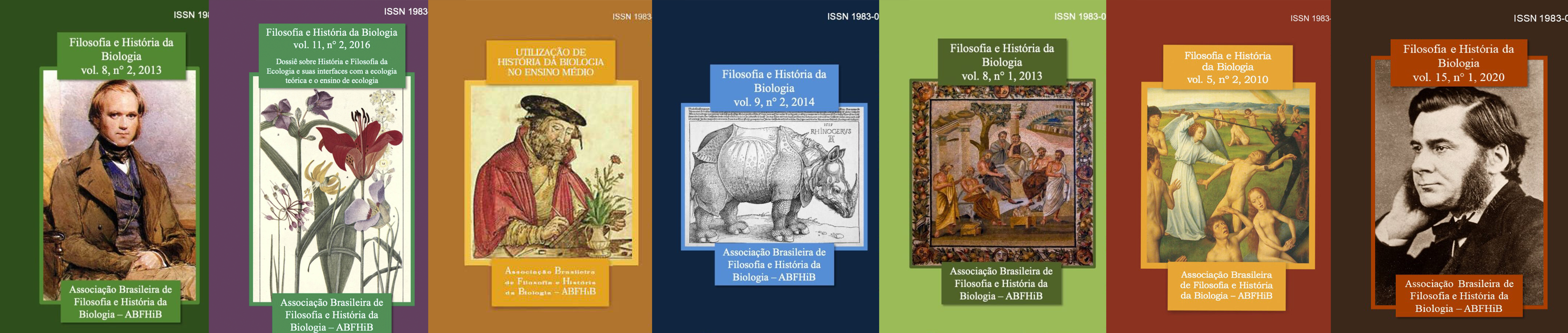
Filosofia e História da Biologia
Philosophy and History of Biology publishes papers resulting from original research regarding philosophy and/or history of biology and its epistemic interfaces, such as history and philosophy of biology and scientific education.
-
Interface Communication Review
The Interface Communication Review has institutional links with the Study Group on New Narratives/GENN of the School of Communications and Arts of the University of São Paulo, and arises from the need to expand the field and area of studies on new perceptions, creations and uses of the concept of narratives.
The magazine's mission is to group, organize and disseminate knowledge and reflections on contemporary narratives, with an emphasis on the fields of Organizational Communication, Public Relations, Publicity and Propaganda, Journalism, Cinema, Tourism, History, Social Sciences and Information Sciences, in addition to the constant interface with the fields of History, Psychology, Anthropology, Sociology, Journalism, Administration, Arts, Literature, Law, Human Rights, Public Policy, Law, Museology, Architecture and Design.
The Editorial Team is composed of the Editorial Committee and the Editorial Board.
-
Revista de Teoria e Pesquisa Econômica
A Revista de Teoria e Pesquisa Econômica foi editada até 1970 pelo Departamento de Economia da Faculdade de Economia, Administração e Contabilidade da Universidade de São Paulo, quando passou a denominar-se Estudos Econômicos.
-
Leviathan (São Paulo)
The main objective of the Revista Leviathan is to offer a plural space for the publication of original works, reviews, research notes, translations and other important materials for the different branches of Political Science.
The journal is aimed at researchers in Political Science, International Relations and related fields, such as Political Philosophy, Sociology, Economics and Statistics. With regard to publications, we do not make any distinction in terms of methodology, approach or institutional affiliation of the authors, and the texts are approved exclusively because of their originality and excellence. Except for occasional translations, all works published in Leviathan are unpublished and represent relevant contributions to the state of the art of Political Science and International Relations.
-
Revista Ingesta
Revista Ingesta is an electronic academic journal, produced by postgraduate students who are also members of the Laboratory of Historical Studies on Drugs and Food (LEHDA), in the History Department of the Faculdade de Filosofia, Letras e Ciências Humanas from Universidade de São Paulo (USP), Brazil. Published semiannually, the journal aims to spread works in this expanding research area and to contribute to its consolidation. We publish articles, book reviews and thematic dossiers in Portuguese, Spanish, and English, produced by postgraduate students and professors in the area of History and related sciences.
-
Revista de Direito Mercantil
A Revista de Direito Mercantil, Industrial, Econômico e Financeiro (“RDM”) foi criada em 1951 em formato impresso e depois teve algumas edições em formato digital. Para visualização de edições anteriores pela Editora Vlex, acesse: https://livros-e-revistas.
vlex.com.br/source/revista- direito-mercantil-industrial- economico-financeiro-11164 ISSN 0102-8049
-
Zi Yue
A Zi Yue é uma revista dedicada aos estudos na área de sinologia. A submissão de trabalhos é feita em formato digital no endereço eletrônico da Revista Zi Yue, observando as regras editoriais disponíveis em em nosso site, aqui. Os artigos são submetidos a pareceristas vinculados ao domínio de conhecimento de cada trabalho.
As opiniões expressas nos artigos assinados são de inteira responsabilidade de seus autores. Todo material incluído nesta revista tem a autorização expressa dos autores ou de seus representantes legais.
-
Revista de Graduação USP
Revista de Graduação USP (Grad+) is an electronic journal that became biannual in 2023. It is under the responsibility of Universidade de São Paulo's Pro-Rectory of Undergraduate Studies. This periodical, which only accepts original articles, is a privileged space for the reflection and sharing of research, pedagogical experiences and teaching practices in the scope of higher education, taking a special interest in undergraduate studies.
Revista de Graduação USP has higher education teachers, graduate students, basic education teachers and educational management professionals, as well as researchers that focus on this theme as its target audience.
-
La Junta (São Paulo)
A La Junta - Revista de Graduação em Espanhol é organizada pelas/os alunas/os de graduação e pós-graduação da área de Língua Espanhola e Literaturas Espanhola e Hispano-Americana do Departamento de Letras Modernas da Faculdade de Filosofia, Letras e Ciências Humanas da Universidade de São Paulo.
-
Revista da Universidade de São Paulo
Revista da Universidade de São Paulo foi publicada de forma irregular entre os anos de 1950 a 1987. A partir de 1989 passou a ser publicada como Revista USP.
-
Organicom
Organicom addresses in depth a special theme, in each edition, organized in the form of a dossier, bringing reflections on trends for the activity and for research in communication.
-

Revista da Biologia
The Revista da Biologia is a scientific journal aimed at the communication of studies of all subjects of Biology, without any costs to the authors and readers. This journal results of an initiative of academics of different brazilian universities that wish to increase the visibility of the works developped by young scientist and undergraduate or graduate students in the different fields of Biology.
We accept submissions in english (preferably) or portuguese, that present original and new discoveries in the fields of Biology, as well as revisions and essays. All publications are submitted to peer-review. The manuscript will be published immediately after the editorial process is finished.
Rev. Biol., ISSN 1984-5154, DOI 10.7594/revbio
For more information, access: http://www.ib.usp.br/revista/
-
Mare Nostrum
Mare Nostrum is a journal about the Ancient Mediterranean that has been published by the Laboratory of Studies on the Roman Empire and the Ancient Mediterranean (University of São Paulo) since 2010.
-
ARA Journal
ARA Journal is dedicated to reflecting on Museums and Heritage, based on a discussion about current times, in compliance with the meaning of the word that names the magazine. Thus, past time, the present and what is expected of the future constitute privileged focuses.
With each issue, the Museum/Heritage Group (GMP) suggests themes to the Editorial Board (EC), made up of national and international experts, who select them. It is also up to the latter to read the Submissions and endorse referees to analyze the material sent, in compliance with the Regulations.
It is divided into two parts: one dedicated to the different ongoing GMP research, called Dossier; the other focuses on the Submissions, both examined by at least two experts, in order to ensure the intended merit.
-
Revista da Tulha
Journal of the Center of Research Support in Performance Sciences (Nap-Cipem), of the Music Department of the Faculty of Philosophy, Sciences and Letters of Ribeirão Preto/University of São Paulo
-
Biblioteca Escolar em Revista
Biblioteca Escolar em Revista é uma revista da Faculdade de Filosofia, Ciências e Letras / USP-Ribeirão Preto que se dedica à divulgação especializada nos estudos sobre história da leitura, biblioteca escolar, práticas de leitura no âmbito escolar, literatura infanto-juvenil e mediação cultural na biblioteca escolar, publicando principalmente pesquisas originais, como também documentos especiais, traduções e resenhas.
-
Humanidades em diálogo
A revista Humanidades em diálogo é editada por alunos de graduação do Programa de Educação Tutorial (PET) da Universidade de São Paulo: PET Filosofia, PET História, PET Ciências Sociais, PET Sociologia Jurídica e PET Direitos. Os objetivos do periódico são contribuir para o aprofundamento do diálogo entre as diferentes disciplinas e campos do saber da área de humanidades, divulgar a produção de conhecimento elaborada por alunos de graduação, bem como proporcionar a estes alunos uma primeira experiência com publicações acadêmicas. Contamos com periodicidade anual e acolhemos trabalhos inéditos produzidos por graduandos da área de humanidades como artigos, críticas e ensaios, ilustrações e demais produções.
-

Revista de Estudos Culturais
A Revista de Estudos Culturais é uma publicação do Programa de Pós-Graduação em Estudos Culturais da Escola de Artes, Ciências e Humanidades da Universidade de São Paulo (EACH/USP). A revista incentiva a submissão de artigos originais e resenhas em todas as vertentes dos Estudos Culturais.
-
Épistémologiques
A revista Épistémologiques foi publicada entre os anos de 2000 e 2002 pelo Departamento de Filosofia da Faculdade de Filosofia, Letras e Ciências Humanas da Universidade de São Paulo.
-
Management & Public Policies Journal
The Management & Public Policy Magazine is intended for the publication of original articles on current topics in public policy management, preferably guided by an interdisciplinary approach. It is a semiannual electronic journal of the School of Arts, Sciences and Humanities of the University of São Paulo (EACH-USP), in association with the Foundation for Administrative Development (FUNDAP). Contributions to the journal can come from different areas of knowledge, in view of the broad scope of public policy management in its multiple dimensions.
The RG&PP does not charge any type of fees or values for submitting or publishing the manuscripts sent for evaluation and, eventually, published in our volumes.
-
Khronos
Khronos – Revista de História da Ciência (Khronos – Journal of the History of Science) is a biannual publication of the CHC – Interunit Centre for the History of Science at the University of São Paulo (founded in 1988) – focused on the history and epistemology of the natural sciences, life sciences, human sciences, technical sciences and related areas. The perspective is interdisciplinary and aims to stimulate interpretative possibilities of scientific and technical knowledge processes in their historical contexts. It publishes original research findings on topics ranging from Antiquity to the 21st century. In addition to these, it welcomes texts from scientists or institutions, as well as unpublished translations, reviews, news of research projects and other topics of interest to historians.
-
Boletim da Faculdade de Filosofia, Ciências e Letras da Universidade de São Paulo. Mineralogia
Boletim. Mineralogia - Faculdade de Filosofia, Ciências e Letras, Universidade de São Paulo
-
Opiniães
“Enfim, cada um o que quer aprova, o senhor sabe: pão ou pães, é questão de opiniães...”
(João Guimarães Rosa, Grande sertão: veredas) -
Revista Geografia Literatura e Arte
The Geography, Literature and Art Magazine is linked to the Department of Geography of the Faculty of Philosophy, Letters and Human Sciences FFLCH) da Universidade de São Paulo (USP).
-

Revista Extraprensa
Extraprensa is a journal written for publishing scientific essays about the Culture and Communication around Brazil and Latin America by compiling themes like cultural diversity, citizenship issues and other popular cultures expressions, including art, alternative media and scientific methods used at these researches.
Papers' Profile used by Extraprensa Journal: articles, research reviews, scientific reports and experts' chronicles.
-
Intelligere
Intelligere, Journal of Intellectual History, is a biannual, electronic, trilingual (Portuguese, Spanish, English) scientific journal dedicated to the studies of Intellectual History and the History of Ideas.
Intelligere publishes original articles, interviews, book reviews, research news in progress, translations, and documentary sources relevant to intellectual history.
-
Boletim do Instituto de Higiene de São Paulo
O Boletim do Instituto de Higiene de São Paulo foi publicado entre anos de 1919 e 1946. O fato de ter surgido no ano seguinte à criação do Laboratório de Hygiene da Faculdade de Medicina indica a importância do Boletim como veículo de comunicação científica, legitimador e divulgador dos ideais do higienismo como matriz fundante da saúde pública no período.
Nos 28 anos em que foi editado, o Boletim apresentou características editoriais que ajudam a elucidar seu papel na legitimação da instituição à qual se vinculava e também na consolidação de uma matriz de entendimento e intervenção sobre e na saúde pública vigente. Cada um dos 88 números editados publicou um artigo, uma separata ou um comentário, e é notável sua regularidade – interrompida apenas nos anos de 1925 e 1926 –, considerando-se as potenciais dificuldades dos processos de criação, estrutura e formação institucional.
A criação do Laboratório de Hygiene em 1918, a mudança da direção do Instituto de Higiene em 1922 para Geraldo de Paula Souza, a formalização da autonomia do Instituto em relação à Faculdade de Medicina, a mudança para a sede em 1931 e a instalação da Faculdade de Saúde Pública em novembro de 1945, entre outros fatos, mostram o dinamismo da institucionalização do campo da saúde pública em São Paulo e a importância que os sujeitos históricos da época davam à comunicação científica, instrumento legitimador da identidade intelectual pretendida.
A maior parte dos trabalhos publicados são produções acadêmicas de diretores, pesquisadores, instrutores, professores e assistentes ligados ao Instituto de Higiene e também – prática comum na época – separatas publicadas em outros periódicos, trabalhos apresentados em congressos e conferências proferidas em ocasiões diversas.
Fonte: Marques, M. C. da C., & Dolci, M. de C. (2016). Boletim e Arquivos: a comunicação científica até a criação da Revista de Saúde Pública . Revista De Saúde Pública, 50, 62. https://doi.org/10.1590/S1518-8787.2016050000115
-

-
Dissenso: revista de estudantes de filosofia
A revista Dissenso foi publicada entre os anos de 1997 e 1999 pelos estudantes do curso de Filosofia, da Faculdade de Filosofia, Letras e Ciências Humanas da Universidade de São Paulo -
Revista LABVERDE
A Revista LABVERDE, criada em 2010 pelo Laboratório LABVERDE, com periodicidade semestral (março-agosto), tem por objetivo divulgar o andamento e resultado das pesquisas científicas, a nível de pós-graduação e promover eventos e encontros científicos, em suas áreas de atuação. Esta decisão editorial de produção somente em suporte digital teve a intenção de tornar a Revista mais ágil, facilitando tanto a colaboração quanto a leitura de pesquisadores, profissionais e demais interessados em temáticas instigantes e abordagens inovadoras na área de Arquitetura Urbanismo e Design.
-
Revista Entrecaminos
A Revista Entrecaminos é uma revista eletrônica fundada em 2013 por discentes e egressos do Programa de Pós-Graduação em Língua Espanhola e Literaturas Espanhola e Hispano-Americana do Departamento de Letras Modernas da Faculdade de Filosofia, Letras e Ciências Humanas (FFLCH) da USP.
-
Língua e Literatura
A revista Língua e Literatura está vinculada à Faculdade de Filosofia, Letras e Ciências Humanas (FFLCH) da Universidade de São Paulo (USP).
-

GIS - Gesture, Image and Sound - Anthropology Journal
GIS – Gesture, Image and Sound – Anthropology Journal is an academic journal that encompasses fields of visual anthropology, music and sound, performance, theater and art.
With the purpose of creating a space for international dialogue involving materials and reflections produced by these fields, we accept publications in Portuguese, Spanish, English and French; in the case of articles published in Portuguese and Spanish, authors should also provide English translations.
The bilingual requirement regarding articles in Spanish and Portuguese is aimed at making known, on a wider scale, production from Latin America and other Portuguese and Spanish-speaking areas.
-
Estudos Japoneses
A revista Estudos Japoneses tem como missão publicar artigos de perfil acadêmico que tratem de temas relativos à Língua, Literatura e Cultura Japonesa, abordados à luz de metodologias científicas. A revista publica artigos em português, inglês, francês, espanhol e japonês.
-
Revista Música
The “Revista Música”, is an academic refereed journal, published by the Graduate Program in Music of the School of Communication and Arts, University of São Paulo (Brazil). Founded in 1990, this journal publishes predominantly original articles, including also other types of significant contributions to the field of research in music (translations, interviews, reviews, scores).
We welcome submissions on any aspect of music research in English, Spanish, and Portuguese. Reviews of books are also welcome. This journal accepts submissions continuously.
Submission guidelines: http://www.revistas.usp.br/revistamusicaShould you have any questions, do not hesitate to contact the editor: revistappgmus@usp.br
-
Acta Fisiátrica
Acta Fisiátrica (ISSN 0104-7795 | e-ISSN 2317-0190) is an open-access scientific journal published quarterly electronically. Acta Fisiátrica is the official publication of the Institute of Physical Medicine and Rehabilitation of the Hospital das Clínicas and the Department of Legal Medicine, Bioethics, Occupational Medicine and Physical Medicine and Rehabilitation of the Faculty of Medicine of the University of São Paulo (IMREA HCFMUSP), being supported by the Faculty of Medicine Foundation Medicine and the Brazilian Association of Physical Medicine and Rehabilitation (ABMFR).
Acta Fisiátrica has no submission, processing, or publication fees and does not generate any economic advantages for the Institute of Physical Medicine and Rehabilitation of the Hospital das Clínicas of the Faculty of Medicine of the University of São Paulo. The institution covers the publishing costs entirely as part of its social and corporate responsibility. To increase the internationalization of the articles published in Acta Fisiátrica, the authors must bear the English translation costs of the accepted manuscript when submitted in Portuguese.
-
Rapsódia
Rapsódia, um almanaque de filosofia e arte que procura mostrar as diferentes manifestações do que se costuma chamar estética.
Nos seus fascículos, ao lado de artigos sobre cinema, poética, literatura, artes plásticas, música, arquitetura e mídia eletrônica - com especial atenção à análise de fenômenos artísticos nacionais - encontram-se entrevistas, traduções, ensaios fotográficos, ficção, poesia, gravuras.
Tendo sido concebida e realizada por pós-graduandos em Filosofia da Universidade de São Paulo, rapsódia procura veicular e reunir pesquisas, contribuindo assim para uma visão de conjunto da nossa produção acadêmica em matéria de filosofia e arte. Mas a invenção artística irrompe de todos os lados, e talvez a maioria não brote em solo universitário.
-

9 Arte (São Paulo)
9ª Arte is a semiannual publication by the Observatório de Histórias em Quadrinhos of the Escola de Comunicações e Artes of Universidade de São Paulo.
-

Revista de Estudios Brasileños
The Revista de Estudios Brasileños (REB) is edited by Ediciones Universidades de Salamanca with the support of the Universidade de São Paulo. The objective of the magazine is to encourage and establish academic-scientific discussions and disseminate ideas and research, constituting a platform for Brazilian studies.
-
Revista Digital de Direito Administrativo
The Administrative Law Digital Review of USP - RDDA (ISSN: 2319-0558) is a digital and free journal created to promote the publication of scientific articles in the field of general and sectoral administrative Law. Articles must aim to evidence the relationship between Law, Public Administration and development. Authors are therefore asked to take into account one of the following questions: how can legal administrative problems harm State and society? How do new institutes and rules of administrative Law contribute to the proper functioning of the Public Administration and, ultimately, improve living conditions?
Articles in english, german, spanish, french and italian may be published are accepted for publication in the original version.
-
Cadernos de Filosofia Alemã: Crítica e Modernidade
Cadernos de Filosofia Alemã: Crítica e Modernidade
University of São Paulo, Brazil
ISSN Printed: 1413-7860
ISSN Online: 2318-9800
Open-Access, Blind Review Periodic
The journal Cadernos de Filosofia Alemã: Crítica e Modernidade, edited by the University of São Paulo, publishes articles, reviews, translations and interviews from national and international scholars in open access since 1996. Our thematic scope comprises not only German Philosophy, but, more generally, reflections concerning modernity, as understood from the theoretical framework provided by the heritage of critical philosophy.
This journal was created by the group FiCeM - Filosofia Crítica e Modernidade (Critical Philosophy and Modernity), a group of scholars of several Brazilian universities, and our Editorial Board gathers scholars from universities in Latin America, United States and Europe.
Currently, we publish articles, reviews and interviews in Portuguese, Spanish, French and English and translations to Portuguese - in volumes released at least every six months and in special Dossiers.
The texts can be submitted at any time in our website (in case of doubt, contact us sending a message to filosofiaalema@usp.br).
-
Revista da Faculdade de Direito, Universidade de São Paulo
A Revista da Faculdade de Direito de São Paulo foi publicada entre os anos de 1893 e 1934. Com a criação da Universidade de São Paulo a Faculdade foi incorporada à USP juntamente com sua revista. A partir de então, passou a ser publicada com o título Revista da Faculdade de Direito da Universidade de São Paulo. -
Cadernos Wittgenstein
Os Cadernos Wittgenstein foram publicados entre os anos de 2000 e 2001 pelo Departamento de Filosofia, da Faculdade de Filosofia, Letras e Ciências e Humanas da Universidade de São Paulo. Em 2016 todos os números publicados foram digitalizados e disponibilizados no Portal de Revistas da USP, pelo Sistema Integrado de Bibliotecas da USP. -
Anagrama
Revista científica interdisciplinar e interinstitucional de graduação publicada pelo MIDIATO - Grupo de Estudos de Linguagem: Práticas Midiáticas do Departamento de Jornalismo e Editoração da Escola de Comunicações e Artes da Universidade de São Paulo - ECA/USP
-
Malala, International Journal of Studies on the Middle East and the Muslim World
Malala - International Journal of Studies on the Middle East and the Muslim World is an on-line journal, a plural publication open to all of those who have original works, directly on or in dialogue with Islam and the Muslim World. The journal was an initiative from the Working Group on the Middle East and the Muslim World (GTOMMM) under the Laboratory for Asian Studies (LEA) at the Faculty of History in the University of São Paulo (DH / FFLCH-USP).
The publication project was born and remains structured around a few ideas: the search for interdisciplinarity in the field of studies on the Middle East and the Muslim World; the search for the definition and affirmation of a field of studies in Brazilian academia (without giving up the international insertion of the debate on Islam, the Middle East and the Muslim World), and an innovative, plural format, accessible to the widest public, thus contributing to scientific dissemination and broadening the debate, seeking greater dialogue with sectors of civil society, the 3rd sector and actors who are often involved with the subject, but outside academia.
In this space, we welcome contributions and debates related to developments in the Middle East in the widest concept (which may also include Northen Africa). As for the Muslim world, we also welcome a broader compression of the meaning - including not only Muslim majority societies in Asia and Africa, but also their non-Muslim minorities - in addition to Muslim minorities in Europe, the Americas, and elsewhere - and their interaction with the West. We also welcome contributions about religion, language, cultural expressions, and/or theoretical issues that fit our orbit.
Malala International Journal of Studies on the Middle East and the Muslim World is a publication which welcomes contributions on a continuous basis in Portuguese, English and Spanish. Calls for papers are issued twice a year, with the possiility of thematic and dossier proposals. Original manuscripts should be submitted in Word format via the journal's website:
-
Autopsy and Case Reports
Autopsy and Case Reports (Autops Case Rep) is an Open Access online medical journal published on continuous flow by the Hospital Universitário of the Universidade de São Paulo (HU USP), under the online ISSN 2236-1960. Since 2018, Autopsy and Case Reports publishes using a continuous publication workflow, allowing the immediate posting of articles in their final form on the journal website as soon as they are ready for publication.
 Accepted articles will be published as Open Access and distributed under the terms of the Creative Commons Attribution License, which permits unrestricted use, distribution, and reproduction in any medium provided the original work is properly cited.
Indexing Bibliographic Databases
Indexing Directories
Archiving and preservation
All articles published are indexed and deposited in full text version for preservation in PubMed Central® (PMC) (https://www.ncbi.nlm.nih.gov/pmc/journals/2811), SciELO Brasil (https://www.scielo.br/autopsy) and at Revistas USP repository of Universidade de São Paulo (http://www.revistas.usp.br/wp/?p=76).
-
Ciência e Filosofia
A revista Ciência e Filosofia foi publicada entre os anos de 1979 e 2008 pelo Departamento de Filosofia da Faculdade de Filosofia, Letras e Ciências Humanas da Universidade de São Paulo. Em 2016 todos os números publicados foram digitalizados e disponibilizados no Portal de Revistas da USP, pelo Sistema Integrado de Bibliotecas da USP. -
Caracol
Caracol is a biannual publication in the field of Spanish Language and Spanish/Spanish-American Literature produced by the Department of Modern Literature of the School of Philosophy, Literature and Human Sciences of the University of São Paulo. It aims to publish original contributions in Spanish/Portuguese, interviews, reviews and, potentially, rare texts of interest to the academic debate in the disciplines of Spanish Literature, Spanish-American Literature, Spanish Language and Translation, being the only magazine in Brazil entirely dedicated to Hispanic Studies. Qualis grade: B1.
-
Boletim de Botânica da Universidade de São Paulo
The journal publishes the results of original scientific research developed by Brazilian and foreign investigators in any field of Botany. -
Caligrama (São Paulo. Online)
A revista Caligrama foi publicada entre os anos de 2005 a 2008 pela Escola de Comunicações e Artes da Universidade de São Paulo -

Via Atlântica
The Via Atlântica journal, a bi-annual peer-reviewed publication by the Graduate Program in Portuguese-speaking Literature Comparative Studies at the University of São Paulo, Brazil, aims to bring scholars from Brazil and abroad the results of investigations carried out by experts in the fields of Lusophone Comparative Studies, Comparative Literature, Literature for Children and Youngsters, Lusophone African Literature, Brazilian Literature, Portuguese Literature and other Lusophone literatures and cultures. It is also part of Via Atlântica’s scope the publication of articles that address the interdisciplinary relations of literature to other art forms and to other fields of knowledge. Every issue of Via Atlântica comprises a leading “Thematic Section” and other eventual sections as “Other Essays”, “Interviews” and “Reviews” of books of interest to Lusophone Comparative Studies and related areas. Via Atlântica is rated in CNPq’s fields of knowledge table as an Other Vernacular Literatures publication (8.02.07.00-6).
-
Boletim IG-USP. Série Didática
O Boletim IG-USP. Série Didática dedicou-se a divulgar pesquisas científicas nas diversas áreas da Geologia, com publicação de artigos científicos originais, criteriosamente avaliados, sobre temática acadêmica e aplicada.
-
Brazilian Journal of Oceanography
To publish scientific contributions on Oceanography including physical oceanography, marine chemistry, marine geology and geophysics, biological oceanography, fisheries and related fields. Since April 2020 it became known as Ocean and Coastal Research
-
Cadernos de Ética e Filosofia Política
A revista Cadernos de Ética e Filosofia Política, cujo número inaugural foi lançado em 1999, tem, ao longo de mais de duas décadas, ininterruptamente e periodicamente publicado artigos dedicados à área de Filosofia. Os Cadernos visam suprir em alguma medida a demanda por textos especializados e que atendam o estado atual da questão para o campo ao qual se destina, fornecendo bibliografia a um público interessado no caráter multifacetado da reflexão sobre a ética e a política. No quadriênio 2021-2024 da Qualis Capes, a revista foi classifica no estrato A2 na área de Filosofia.
As questões relativas ao direito, à história, à religião e às artes não raro são por elas incorporadas, convertendo a um só tempo em sua matéria de investigação e seu cenário de intervenção. É esse caráter abrangente da ética e da filosofia política que lhes concede a virtude da vivacidade. Os Cadernos sempre procuraram corresponder e promover essa virtude, veiculando sobretudo a produção teórica discente, sem distinguir correntes ideológicas, linhas filosóficas ou áreas de saber incluídas nas mais diversas manifestações de reflexão.
Aqui se encontrarão artigos, ensaios, resumos de teses e dissertações, resenhas, traduções de trechos de obras e de pequenas obras. Todos os trabalhos, de recepção dos artigos, envio para pareceristas, revisão, editoração e publicação são realizados pela equipe editorial, que se reúne regularmente e toma suas decisões editoriais de modo autônomo.
A revista é editada em meio eletrônico, pelo sistema OJS, o que resulta em um ganho substancial de qualidade, pois facilita o acesso e a difusão dos textos. Somando-se a isso, contamos com um vasto corpo de pareceristas especializados nos temas, correntes filosóficas e autores enfocados pelos artigos, o que torna mais democrática a escolha dos textos destinados à publicação.
Além disso, os Cadernos utilizam o sistema de avaliação na modalidade double-blind review, nos quais todos os manuscritos enviados passam por ao menos dois avaliadores. De 2017 até o presente momento, a revista somente recebe artigos no prazo aberto de chamadas, noticiadas nesse site e em outros meios. Finalmente, desde 2015 os Cadernos têm promovido eventos na área, buscando contribuir para o debate sobre a pesquisa filosófico-política no Brasil, bem como tem editado dossiês temáticos, conduzidos, conjuntamente com o corpo editorial da revista, com editores e editoras convidados.
Convidamos os/as estudantes de pós-graduação em filosofia e pesquisadores/as interessados/as em publicar seus trabalhos a colaborar conosco, ajudando-nos a diminuir assim a distância entre a pesquisa individual e o diálogo aberto com autores e críticos.
-
Alterjor Journal
Alterjor is a scientific journal issued semiannualy by the Research Group Alterjor (ECA/USP), wich deals with popular and alternative Journalism. The popular Journalism is defined by the journalistic practices that occurs among the organised social and popular movements, includind the Third Sector, whose objetives tackles the fostering of institutional organisations and the spread of the leadership of non-hegemonic social segments. The Alternative Journalism is dealed by the experiences of Journalism through the several media and also tackle the fostering of public debate, described as Popoular Journalism. Furthermore, the Alterjor Journal invites the reader to think the achievement of the communication media democratisation for every segments of the society.
-
Significação: Journal of Audiovisual Culture
Significação - Revista de Cultura Audiovisual publishes articles dedicated to the study of audiovisual media and digital systems, considering them in their diversity of practices and ideas that involve specific processes of reflection, creation, production and dissemination.
-
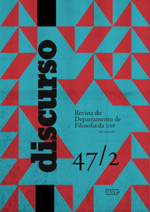
Discurso
A revista Discurso, órgão oficial do Departamento de Filosofia da USP, surgiu em 1970. Quando atravessava a mais difícil fase de sua história, atingido duramente pela violência da ditadura, este Departamento extraiu da ameaça de seu desaparecimento a força e a coragem para criar um espaço de expressão.
A revista propôs-se a veicular não apenas a produção teórica de seu corpo docente, mas também as mais diversas manifestações de reflexão sobre a cultura, sem distinguir correntes ideológicas, linhas filosóficas ou áreas do saber. Assim se pretendia garantir o pluralismo e a liberdade, numa época de obscurantismo.
ATENÇÃO
As submissões à revista Discurso encontram-se suspensas para devido tratamento dos próximos números. Pedimos aos nossos usuários e autores que acompanhem nosso quadro de notícias a fim de tomarem conhecimento sobre futuras datas de submissão.
Att.,
Equipe Editorial
-
Letras Clássicas

Letras Clássicas de http://www.revistas.usp.br/letrasclassicas/index está licenciado com uma Licença Creative Commons - Atribuição-CompartilhaIgual 4.0 Internacional. -
Arquivos da Faculdade de Higiene e Saúde Pública da Universidade de São Paulo
Os Arquivos da Faculdade de Higiene e Saúde Pública da Universidade de São Paulo foram publicados entre os anos de 1947 e 1966. -
RuMoRes
RuMoRes – Online Magazine on Communication, Language and Media is a semiannual scientific journal edited by MidiAto – Group of Study on Language and Media Practices at the School of Communication and Arts of the University of São Paulo (ECA-USP) dedicated to publishing scientific articles, critical reviews and interviews that contribute to the debate on communication, culture, media and language. It is ranked as a B1 magazine on the Coordination for the Improvement of Higher Education Personnel (CAPES) Qualis and accepts the submission of original and unpublished works (written individually or collectively) from authors of a minimum academic level of PhD or doctoral students enrolled on higher education institutions. The submission are continuous and the texts should be formatted on Word using 12 TNR, 1,5 line spacing and following the directions found at Submissions. The magazine finds support at the Postgraduate Program in Audiovisual Media and Processes and at the Postgraduate Program in Communication Sciences of ECA-USP. Access our website at www.rumores.usp.br and for other information write to rumores@usp.br.
-
Estudos Semióticos
Estudos Semióticos (ISSN 1980-4016) is an open-access online publication of the Graduate Program in Semiotics and General Linguistics at the University of São Paulo. It publishes papers and book reports in Semiotics, as well as in adjoining fields. Works dealing with signs, texts, discourses and social practices are welcome, as long as they are original, unpublished papers which establish a dialogue with semiotic theories. Manuscripts are received in five languages: Portuguese, French, English, Spanish, and Italian.
Each annual volume contains three issues: two thematic numbers and one varia. Occasionally, a special dossier may be added as an additional issue to the other three.
At the last evaluation by the Brazilian research organism CAPES, Estudos Semióticos was ranked at the A2 layer (QUALIS-CAPES).
-
InCID: Revista de Ciência da Informação e Documentação
InCID: Revista de Ciência da Informação e Documentação is an open-access scientific journal published by USP-Ribeirão, dedicated to specialized dissemination of Information Science. It publishes mainly original research, along with special documents, translations, and reviews
-
Clinical and Laboratorial Research in Dentistry
Clinical and Laboratorial Research in Dentistry - CLRD, is a peer-reviewed quarterly journal, publishing original research, clinical trials and review articles, editorials, and commentaries related to all areas of Dentistry and those interested in these fields. The journal is dedicated to the dissemination of knowledge and information relevant to dentistry. Submitted manuscripts will be evaluated considering originality, relevance and methodology. The content submitted must not be under consideration elsewhere.
The journal is indexed in the following databases:


Visit and follow the news on our website!
https://www.facebook.com/clresearchindentistry

-
Journal of Information Systems and Technology Management
The Journal of Information Systems and Technology Management - JISTEM has the mission to publish relevant research to the management of technology and information systems in organizations and society.
-
Agrária (São Paulo. Online)
A revista Agrária tem como proposta a construção de um canal de debate em torno da questão agrária, em uma perspectiva crítica, com o compromisso de contribuir para a compreensão de seus fundamentos e suas diferentes formas de manifestações territoriais.
Nesse sentido a revista debate temas atuais apresentados geralmente sob a forma de dossiês, com contribuições de pesquisadores nacionais e internacionais. A revista possui as seguintes seções: Artigos, Teoria em Debate e Resenhas.
-
MATRIZes
MATRIZes is a journal for the publication of scientific production whose object of study is communication. It hosts theoretical works, analysis experiences and conceptual formulations on communicative processes, means, mediations and emergencies of interactions in the contemporary generalized information society. It is a journal open to reflections on the historical transformations of mediations in culture; on the production of languages and their interfaces; about the socio-political implications of the activities implemented, as well as their cognitive consequences. To this end, it preserves the inter and transdisciplinary horizon of the theoretical and methodological contributions of communicational thinking. It is expected, therefore, to resize historical knowledge and traditions that contribute to define, map and conceptually explore communicative events, reviving the commitments that are also part of the history of PPGCOM-ECA-USP. At the limit, the need to create a space for the construction of a critical and consequent theory of communication study practices is evident.
-
Anais do Museu Paulista: História e Cultura Material
Publish theoretical and monographic articles that are based on social practices mediated by materiality and addressed as historical, museological, and conservation issues.
-
Brazilian Journal of Latin American Studies
A Brazilian Journal of Latin American Studies (Cadernos Prolam/USP) é uma revista científica especializada em difundir conclusões de pesquisa, análises e interpretações, bem como pensamento e teorias sobre a América Latina. Criada em 2002, pelo Programa de Pós-graduação Integração da América Latina (PROLAM/USP), desde sua criação é uma revista voltada para autores e público de nível de pós-graduação. Na fase inicial o foco das publicações da BJLAS era as relações internacionais. Com o passar dos anos, o periódico ampliou seu universo disciplinar e temático, e hoje publica trabalhos nos diversos campos das humanidades, artes e ciências sociais.
Assim, tem como eixo organizador incluir temáticas (a) de impacto regional para a América Latina ou (b) trabalhos com metodologias comparativas sobre dois ou mais países deste continente.
Considera-se que os manuscritos devem contribuir de modo significativo ao avanço do conhecimento científico em temáticas sensíveis à América Latina, por este motivo, as propostas publicadas são elaboradas por autores com nível de pós-graduação. As problemáticas que tratam de América Latina exigem perspectivas transdisciplinares com abordagens sobre tópicos transversais em questões sociais, políticas, econômicas, jurídicas, históricas, culturais, artísticas, de comunicação social.
Finalmente, a BJLAS tem interesse em divulgar resenhas de livros recentemente publicados ou de obras de grande relevância para a região, como clássicos do pensamento latino-americano, e aceita também críticas de arte ou ensaios de qualidade.
-
Resenhas do Instituto de Matemática e Estatística da Universidade de São Paulo
A revista Resenhas do Instituto de Matemática e Estatística da Universidade de São Paulo foi publicada entre os anos de 1993 e 2005. A partir desta data continuou a ser publicada com o título São Paulo Journal of Mathematical Sciences -
Arquivos de Zoologia
Arquivos de Zoologia, formerly “Arquivos de Zoologia do Estado de São Paulo”, v.1 (1940) - v.14, (1966), it is a traditional journal in the field of Zoology in Brazil.
The journal aims to publish original scientific papers in the areas of systematics, paleontology, evolutionary biology, ecology, taxonomy, anatomy, behavior, functional morphology, molecular biology, ontogeny, faunistic studies, biogeography, and history of museums and collections of natural history.
Published by the Museu de Zoologia da Universidade de São Paulo (MZUSP), the journal is indexed in the main reference databases and displayed every 6 (six) months.
-
Pesquisa em Educação Ambiental
Publishing original papers related to environmental education research, contributing to the producation of scientific knowledge in this area and to improve practices related to environmental education.
-
Pesquisa Odontológica Brasileira
To publish basic and applied research works, as well as articles on current themes and updated information in oral research. -
Psicologia USP
To publish original works which may contribute to the knowledge and development of Psychology and related fields, such as theoretical articles and essays emphasizing some classical issues such as Memory, Family, Conscience, Unconscious, and Alterity.
-
Boletim IGA
To divulgate geoscientific research by means of the publication of carefully analysed, original papers concerning academic and applied themes in the various Geology areas. -
Revista Brasileira de Ciências Farmacêuticas
A origem da Revista Brasileira de Ciências Farmacêuticas / Brazilian Journal of Pharmaceutical Sciences remonta aos “Anais de Farmácia e Odontologia da USP”, periódico iniciado em 1939. Em 1963, com o vol.1, teve início a “Revista da Faculdade de Farmácia e Bioquímica da Universidade de São Paulo”, que, em 1970, recebeu o título de “Revista de Farmácia e Bioquímica da Universidade de São Paulo”. Em 1999, foi totalmente reformulada, recebendo a denominação de Revista Brasileira de Ciências Farmacêuticas/Brazilian Journal of Pharmaceutical Sciences.
OBS.: O revista mudou de título em 2009 para Brazilian Journal of Pharmaceutical Sciences
-
Revista de Estudos Orientais
A Revista Estudos Orientais é uma publicação do Departamento de Letras Orientais da Faculdade de Filosofia, Letras e Ciências Humanas da Universidade de São Paulo. As linhas de pesquisa da Revista concentram-se nos estudos linguísticos, literários e culturais das áreas constituintes do Departamento: Árabe, Armênio, Chinês, Coreano, Hebraico, Japonês e Russo. Outras áreas ligadas aos estudos asiáticos e eslavos completam esse escopo. São aceitos para publicação artigos, resenhas, traduções e entrevistas relacionadas às áreas de pesquisa mencionadas.
A Revista de Estudos Orientais está retomando as suas atividades com uma nova equipe, e cumprindo com todas as exigências acadêmicas necessárias para uma rápida atualização e evolução do seu conceito.
-
Celeuma
Celeuma é uma palavra estranha e um pouco esquecida. Ela significa “discussão intensa”, “agitação ruidosa”. Não são brigas ou gritos, contudo, que o leitor deve esperar do material da nova revista, publicação eletrônica que terá três números por ano e que agora é lançada nas comemorações dos 20 anos do Centro Universitário Maria Antonia vinculado à Pró-Reitoria de Cultura e Extensão Universitária da Universidade de São Paulo.
O tema de Celeuma são as artes na atualidade: a crítica, as teorias da ficção e da narrativa, os impactos da tecnologia, a mistura cada vez mais complexa entre as diferentes linguagens. E aí, em seus assuntos, se confundem estranhezas e esquecimentos, discussões e ruídos. É um campo vasto de pesquisa e conversa, para alguns fundamental e constitutivo, para outros insignificante e instrumentalizável, e, até por essa disparidade de percepções, cada vez mais urgente e necessário. Daí esse nome: este o seu propósito.
-
Cadernos CERU
The Journal aims to divulge results of investigations from Brazilian and Foreign researchers in the several fields of Social Science.
-
Caderno de Estudos
The mission of Cadernos de Estudos is to disseminate relevant scientific production in Accountancy, Controllership, Actuarial Science and Finance, produced by Brazilian and international faculty, researchers, students and professionals, exclusively selected based on quality and actual contribution to knowledge development in this field, subject to blind review. -
Revista da Faculdade de Medicina Veterinária, Universidade de São Paulo
1938-1971: A Revista da Faculdade de Medicina Veterinária é publicada sob a forma de fascículos que serão reunidos em volume e não tem data certa de aparecimento.
Título abreviado: Rev. Fac. Med. Vet.
ISSN: 0301-7273
eISSN: 2318-5066- Continua como: Revista da Faculdade de Medicina Veterinária e Zootecnia da Universidade de São Paulo (1972-1989).
ISSN: 0303-7525
e-ISSN: 2318-3659
- Continua como: Revista da Faculdade de Medicina Veterinária e Zootecnia da Universidade de São Paulo (1972-1989).
-
Journal of Ancient Philosophy
The Journal of Ancient Philosophy is an e-journal published by the Department of Philosophy of the Universidade de São Paulo (USP, Brazil). It was founded in 2007 and publishes articles, reviews and textual notes on ancient Greek and Roman philosophy, as well as translations of classical texts into Portuguese or Spanish. The journal also intends to provide information about Latin American symposia, meetings and conferences on ancient Greek and Roman philosophy. It is published twice a year, in May and October.
The purposes for which the Journal of Ancient Philosophy was created are to foster classical studies in Latin America, providing scholars a vehicle for the publication of researches and discussions, and to promote international dialogue across different languages and approaches. It is open to all scholars worldwide to submit contributions for inclusion in this Journal. The accepted languages are: Portuguese, Spanish, English, French, German and Italian. Every contribution is evaluated by a double-blind, peer-review process.
This Journal considers plagiarism as a very serious offense against academic research, and consequently will take all measures to protect its publications from it, aiming at ensuring originality in any paper published by this Journal. This Journal will not condone plagiarism under any circumstances. Please report to www.fapesp.br/6566 for a theoretical assessment of plagiarism, and www.fapesp.br/6574 for practical and general information on sound academic practices of quotation and publication. We fully agree with the proposals put forward by Fapesp in these documents.
Likewise, submissions that have their origin in artificial intelligence (AI) programs are not considered academically relevant. As we know, such programs are not capable of bringing original contributions to the field of study to which the Revista de Filosofia Antiga is dedicated. All contributions originating from AI tools will be disregarded.
Abstracted / Indexed in: Philosopher's Index, Catálogo LatinIndex, EBSCOhost, Portal de Periódicos da CAPES, Qualis CAPES

This work is licensed under a Creative Commons Attribution-NoDerivatives 4.0 International License. -
Teresa
Teresa – Revista de literatura brasileira (Qualis B2), uma publicação da área de Literatura Brasileira da Universidade de São Paulo (USP), criada em 2000, tem como objetivo publicar pesquisas e investigações, abordando a área de Literatura Brasileira em perspectivas originais e avançadas, desenvolvidas por pesquisadores no Brasil e no exterior.
-
Educação e Pesquisa
Education and Research is a publication of the School of Education of the University of São Paulo – FEUSP. Since 2018, it is published in a single issue per year. It publishes original articles on education in Portuguese, Spanish and English. The articles result from theoretical or empirical research or literature reviews characterized by a consistent dialogue with the educational field and the area of study to which they are affiliated, by detailing of conceptual and methodological work, by the explanation of analytical processes and by the relevance of their contribution to the development of knowledge in Education. -
Revista de Saúde Pública
To publish and divulge scientific production on subjects of relevance to Public Health.
-
Estudos Avançados
The Reviews guiding proposal is to leverage knowledge and critique for social progress, both for Brazilians and for other developing peoples.
-
Revista Aspas
Journal of Graduate Program in Performing Arts - University of São Paulo
-
Revista do Instituto de Estudos Brasileiros
The University of São Paulo's Brazilian Studies Institute is a multidisciplinary centre for education, research and documentation on the culture of the nation. With a staff of scholars from various backgrounds, the centre offers graduate, post-graduate, diffusion and extension courses, as well as orientation in scientific initiation, master and post-doctoral studies. To reflect on Brazilian society through the articulation of multiple fields of knowledge was the challenge at the heart of the IEB's foundation in 1962, by Sergio Buarque de Holanda, and it has motivated the production of a wealth of original research ever since, some of which has found its outlet in its Revista (Journal). Created in 1966, the Journal of the Brazilian Studies Institute - USP was published without interruption until 1997. Given the very nature of the institute, the Journal always followed a multidisciplinary approach, embracing different trends and lines of research and opening its pages to researchers from an enormous diversity of institutions. In many fields of knowledge, the Journal played a part in the drive to renew Brazilian studies that began in the 1960s, serving as one of the key outlets for the diffusion of original scientific research from Brazilian universities and their post-graduate programs between 1970 and 1990. It was also one of the vehicles for the divulgation of the IEB's vast archive, which today includes the personal collections of such fundamental names in Brazilian culture as Mário de Andrade, Graciliano Ramos, Guimarães Rosa, Osman Lins, Alberto Lamego, Yan de Almeida Prado, José Honório Rodrigues, Caio Prado Jr., Pierre Monbeig, Anita Malfatti, Francisco Mignone and Camargo Guarnieri. The IEB Journal, a forum for debate devoted to incorporating a multiplicity of themes, schools of thought and lines of research, was resumed in September 2006 with the proposal of integrating the various branches of the Humanities, accepting the challenge of critically thinking Brazil and facilitating dialogue between different fields of knowledge - though without compromising on the specificities and technical rigor demanded by the specialist reader. Its goal is, therefore, to present studies of such theoretical and methodological relevance and significance that they can and should transcend compartmentalization. Furthermore, it is with that same objective of promoting debates in which knowledge from disparate origins can meet and interact that the IEB Journal opens its pages to both the analysis of problems facing contemporary Brazil and to reflections that investigate theoretical dimensions of Brazilian studies. To this end, it assumes the challenge of molding itself into an agglutinative - but not harmonizing - forum for the countless frontiers of knowledge.
-
Fisioterapia e Pesquisa
Basic Information
The Physical Therapy and Research Journal (Revista de Fisioterapia e Pesquisa) (ISSN 1809-2950), continuation of the University of São Paulo Physiotherapy Journal (Revista de Fisioterapia da Universidade de São Paulo) (ISSN 1413-7879), is a scientific journal created in 1994, whose mission is to disseminate scientific production and knowledge in the field of physiotherapy and rehabilitation and related disciplines. In aspects related to basic, clinical, and applied research.
Frequency: quarterly
INDEXING SOURCES
SCIELO- Scientific Electronic Library Online
BVS – Biblioteca Virtual em Saúde (Virtual Health Library)
CINAH – Cumulative Index to Nursing and Allied Health Literature
LILACS – Latin American and Caribbean Health Sciences
LATINDEX – Sistema Regional (Regional System)
Sport DISCUS
Intelectual property
All journal content, except where identified, is licensed under a Creative Commons License of the type attributed BY
Sponsors
The publication receives funding from:
AGUIA – USP Agency for Academic Information Management
FMUSP – USP School of Medicine
CREFITO - Regional Council of Physiotherapy and Occupational Therapy
-
Revista de Italianística
Revista de Italianística is a journal of the Graduate Program in Italian Language, Literature, and Culture at the University of São Paulo. Every six months this journal publishes issues dedicated to linguistic studies and literary studies to disseminate the scientific production of national and foreign researchers associated with the area of Italianistics.
-

-
Revista do Museu de Arqueologia e Etnologia
The Revista do Museu de Arqueologia e Etnologia appeared in 1991, replacing three extinct titles due to the merger of research institutions in the areas of Archaeology and Ethnology: Revista Dédalo, Revista de Pré-História e Revista do Museu Paulista. The Revista do Museu de Arqueologia e Etnologia is an academic journal for the publication of works on Archaeology, Ethnology and Museology, and in 2016, the Revista de Arqueologia e Etnologia became on-line only, semiannual and all previous printed numbers were digitized and made available in open access through Portal de Revistas USP Site. -
The São Paulo Journal of Mathematical Sciences
To publish high-level papers in all areas of mathematics, statistics and computer science.
-
Boletim IG-USP. Publicação Especial
O Boletim IG-USP. Publicação Especial teve como objetivo publicar assuntos temáticos diversos, abrangendo revisões que demandam trabalhos mais longos e as contribuições científicas que fazem parte de eventos.
-
Journal of Human Growth and Development
Journal of Human Growth and Development (JHGD) is an online journal that provides access to high quality and open access and free publication. Was founded in 1991 by Professor Arnaldo Augusto Franco de Siqueira. link: http://revistas.marilia.unesp.br/index.php/jhgd
-
Signos do Consumo
The journal Signos do Consumo specializes in studies on marketing and advertising, public relations, and market and consumption phenomena. Its mission is creating a forum for the exchange of knowledge in a network, through scientific articles and book reviews, for the national and international scientific community interested in disseminating the results of its researches, essays, and reflections concerning studies in the area of Communication. Article languages for publication: Portuguese, Spanish, and English.
ISSN: 1984-5057
-
Literartes
LITERARTES é revista do grupo de estudos Produções Literárias para crianças e jovens da área de Literatura Infantil e Juvenil da FFLCH/USP. Publicação periódica - em princípio, semestral - que visa a propiciar um espaço de reflexões sobre a arte literária e os diálogos que vem estabelecendo com outras linguagens, outras artes e suportes.
-
Revista Crioula
A Revista Crioula é uma publicação científica dos alunos de pós-graduação da Área de Estudos Comparados de Literaturas de Língua Portuguesa do Departamento de Letras Clássicas e Vernáculas da Universidade de São Paulo (ECLLP - DLCV - FFLCH/USP).
O periódico promove a difusão da produção científica dos alunos e ex-alunos do Programa de Pós-Graduação em Estudos Comparados de Literaturas de Língua Portuguesa da USP, bem como de pesquisadores de outras universidades do Brasil e do exterior.
-

Novos Olhares
Novos Olhares – Revista de Estudos Sobre Práticas de Recepção a Produtos Midiáticos, foi criada em 1998 pelo Grupo de Estudos Sobre Práticas de Recepção a Produtos Mediáticos, coordenado por Mauro Wilton de Sousa, Professor Titular da Escola de Comunicações e Artes da Universidade de São Paulo, que se tornou seu editor. Até 2007, quando sua publicação foi descontinuada, a revista contou com 20 edições impressas, tornando-se a principal publicação do país no campo dos Estudos Culturais e contando com a contribuição de renomados pesquisadores nacionais e internacionais.
A partir de 2012, a revista ressurgiu como publicação online vinculada ao MidiaSon: Grupo de Estudos e Produção em Mídia Sonora, da Escola de Comunicações e Artes da Universidade de São Paulo. Eduardo Vicente, Professor Associado da instituição, assumiu, desde então, a função de editor da publicação, que teve o seu Comitê Editorial completamente renovado. Para a retomada, optou-se pela mudança na numeração da revista, que foi reiniciada no formato volume/número. Assim, as edições de 2012 tornaram-se, respectivamente, os números 1 e 2 do Volumes 1.
Desde 2013, a revista é hospedada no Portal de Periódicos da Universidade de São Paulo no endereço www.revistas.usp.br/novosolhares. Além de todas as edições online, são disponibilizadas também as versões digitalizadas das 20 edições impressas da revista que, além de trazer contribuições de autores como Bernard Miège, Lorenzo Vilches, Jesús Martín-Barbero e Antonio Fausto Neto, entre outros, reúnem entrevistas de pesquisadores como Octávio Ianni, Renato Ortiz, Sérgio Adorno, Eduardo Peñuela-Canizal, Arlindo Machado e Maria Lourdes Motter, entre outros.
A partir de 2025, a Novos Olhares passou a ser publicada em fluxo contínuo, com uma única edição/volume anual.
A revista publica, em suas edições, contribuições voltadas a um amplo leque de temáticas dentro da área de Comunicação. Sem abandonar sua vocação inicial, ligada às praticas de recepção, o periódico tem se tornado uma referência também na publicação de trabalhos voltados a diferentes aspectos da produção audiovisual, com destaque para o rádio, o podcast e a música popular industrializada.
-
Non Plus
Revista da Área do Francês da FFLCH – USP
A Non Plus é uma publicação eletrônica semestral do programa de Pós-Graduação em Estudos Linguísticos, Literários e Tradutológicos em Francês, do Departamento de Letras Modernas, da Faculdade de Filosofia, Letras e Ciências Humanas(FFLCH) da Universidade de São Paulo(USP).
A Non Plus publica artigos de caráter teórico que contemplem áreas cujo objeto de estudo seja a linguagem em suas diversas manifestações, tais como os segmentos de Didática, Estudos Literários, Língua e Tradução. Além disso, traz Resenhas de teses e dissertações defendidas relativas às áreas mencionadas, no intuito de difundir a produção acadêmica dedicada às Letras francesas no Brasil.
-
Paisagem e Ambiente
To present and discuss themes related to Open Spaces and Environmental studies, to spread the Brazilian and International scientific and professional production.
-
Plural
Plural is a biannual electronic academic publication, coordinated and edited by scholars from the Graduate Program in Sociology at the Faculty of Philosophy, Languages and Humanities of the University of São Paulo (FFLCH-USP), with Qualis B2 classification in the 2013-2016 quadrennium.
-
Acolhendo a Alfabetização nos Países de Língua Portuguesa
Publish unprecedented articles, resulting from reseach, containing relevant information on the countries whose official language is Portuguese, so that the knowledge about what is happening in education in those places can be accessible to the comunity interested in literacy. -
Pandaemonium Germanicum
The journal Pandaemonium Germanicum, published since 1997 by the German Area of the Department of Modern Letters of the FFLCH/USP and the Post-Graduate Program in German Language and Literature, comprehends itself as a forum of academic discussion in the Germanistic field covering: German language literature, compared literature and cultural studies related to German-speaking countries, German linguistics, applied and contrastive linguistics (Portuguese/German), teaching of German as a foreign/additional language, and studies on translation logic.
Pandaemonium seeks not only to disseminate studies by Germanists from Brazil and abroad, but also to contribute to the dialogue between German studies, other languages and literatures as well as further fields of knowledge.
Pandaemonium is an open access journal published semi-annually until 2015 and three times a year from 2016 on. The manuscripts are submitted to double blind peer review. In case of divergent opinions, a third opinion is sought for. Accepted articles are throroughly edited before publishing.
-
Anais da Escola Superior de Agricultura Luiz de Queiroz
Publish original articles that contribute to the scientific development of Agricultural Sciences. -
ARS
Ars journal is a publication from the Graduate Program in Visual Arts of the School of Communication and Arts (ECA), University of São Paulo (USP). It assembles relevant works on the debate about art produced in- and outside the university. Moreover, it proposes a broad approach according to the requirements of contemporary artistic production. Concerned with a compreehensive range of issues, the journal aims to contribute to the cultural debate outside the barriers of the University and to question perspectives of art in the contemporary context. At the same time, it values the theoretical contribution of ore traditional disciplines, areas such as Philosophy, Aesthetics, and History of Art, although often confronting or pointing out the limits of this tradition in the face of the challenges of contemporary challenges. Ars focuses on running, alongside the works of artists, critics, art historians and graduate students of ECA’s Department of Visual Arts, collaborations of researchers, artists, intellectuals and other professionals in the artistic field, favoring experimentation and theoretical specialized research.
Ars currently runs an eletronic issue (ISSN 2178-0447).
The journal is indexed in SciELO, LatinIndex, DOAJ, and Portal de Revistas da USP.
Rating Qualis/Capes for Arts/Music: A1
-
África
Launched in January 1978, the journal Africa is an international and bilingual periodical of the Center for African Studies at the Faculty of Philosophy, Letters, and Human Sciences of the University of São Paulo (CEA FFLCH-USP).
The journal Africa, published annually and on a rolling basis, contributes to the dissemination of studies about the African continent. It accepts original works, reviews, essays, reading notes, articles, or news related to African, Afro-Brazilian, and the broader Black diaspora realities, in the fields of sociology, anthropology, political science, international relations, geopolitics, history, literature, music, and health.
-
Boletim do Instituto Oceanografico
O Boletim do Instituto Oceanográfico destinou-se a publicar trabalhos originais e de real valor científico, voltados à oceanografia e tecnologia dos produtos do mar e de outras ciências correlatas.
-

Pós. Revista do Programa de Pós-Graduação em Arquitetura e Urbanismo da FAUUSP
Pós- is an international scientific journal, refereed and indexed, administered by the Graduate Program of the Faculty of Architecture and Urbanism of the University of São Paulo - FAUUSP. Its objective is to publish national and international research results through unpublished articles reviewed by the double blind peer review system and thus contribute to the dissemination of scientific production developed in the various areas related to architecture, urban planning and design.
-

GEOUSP Espaço e Tempo (Online)
Journal of the Post-graduation Program in Human Geography and the Post-graduation Program in Physical Geography
Department of Geography
Faculty of Philosophy, Letters and Human Sciences
University of Sao Paulo
Brazil
ISSN: 2179-0892 -
Boletim IG-USP. Série Científica
To divulgate geoscientific research by means of the publication of carefully analysed, original papers concerning academic and applied themes in the various Geology areas. -
Brazilian Journal of Veterinary Research and Animal Science
The Brazilian Journal of Veterinary Research and Animal Science (BJVRAS), eISSN 1678-4456, is continous published, exclusively online, and in English. The BJVRAS is linked to the School of Veterinary Medicine and Animal Science, University of São Paulo (SP, Brazil), being supported by the Veterinary Medicine Foundation (FUMVET). The BJVRAS is intended to publish scientific studies on veterinary medicine and related sciences.
-
Cadernos de Psicologia Social do Trabalho
The Journal of Social Psychology of Work (JSPW) is an on-line, open access, interdisciplinary academic journal which operates in a Continuous Article Publishing (CAP) mode. It aims at promoting scientific production in the fields of Work and Organizational Processes from the Social Psychology perspective, as well as from other fields in humanities and social sciences which fall within the scope of critical perspectives. Priority will be given to contributions that present reflections on concrete situations and propositions that advance in transformation processes towards health promotion and the guarantee of workers' rights. In this sense, Work is considered a central category as well as the Workers’ point of view, marking an epistemological and ethical-political difference from the managerial approaches which consider workers as human resources. This journal welcomes articles based on diverse theoretical readings, original and unpublished, in the following formats: research, intervention and experience reports, as well as theoretical essays. Literature reviews are only accepted when used to support a consistent theoretical discussion. After consultation with the editors, book reviews, translations and interviews may be accepted. The journal’s target audience includes students, academics, researchers, academics and professionals from Psychology and related areas interested in the field of Work and Organizational Processes.
ISSN 1981-0490
cpst@ups.br
-
Revista CPC
The Revista CPC is a scientific journal published by the Cultural Preservation Center of the University of São Paulo, a body of the Office of the Provost for Culture and Extension.The journal focuses on the discussion and reflection of issues related to cultural heritage in its many aspects: heritage preservation; identification and documentation of cultural assets; heritage education; cultural references and social memory; museum institutions; archives, memory and information centers; collections and holdings; conservation and restoration. Published semiannually, the journal features original articles, reviews, interviews, reports, and testimonials. Each year, it publishes a special themed edition, such as a Dossier. Since its creation in 2005, Revista CPC has been regularly dedicated to promoting research and disseminating knowledge in the field of cultural heritage. Published exclusively in digital format, it became part of the USP Journal Portal in 2014.The metadata of Revista CPC is indexed in: Latindex - Sistema Regional de Información en Línea para Revistas Científicas de América Latina, el Caribe, España y Portugal; Periódicos CAPES - Portal of Journals of the Coordination for the Improvement of Higher Education Personnel (CAPES/MEC); Journals for Free - Directory of Open Access Journals; LivRe - Open Access Journals (CNEN-MCTIC); Sociology Source Ultimate - Ebsco. Registered in Diadorim -Directory of Editorial Policies of Brazilian Scientific Journals. Member of the collaborative network LatinRev - Red Latinoamericana de Revistas Académicas en Ciencias Socialies y Humanidades (FLACSO ARGENTINA).
A2 classification by the Qualis Journals evaluation system – Sucupira Platform/CAPES Quadrennial 2017-2020.
-

Economia Aplicada
The aims of the Journal is to publish papers with economic analysis applied to specific problems of interest either to public or private sector, especially with quantitative results bringing theory and reality closer.
-
Estudos Econômicos (São Paulo)
O periódico Estudos Econômicos foi criado em 1971 por iniciativa do Departamento de Economia da Faculdade de Economia, Administração, Contabilidade e Atuária da Universidade de São Paulo.
Publica trabalhos inéditos com contribuição original em qualquer área da Economia, comprometendo-se com padrões acadêmicos de excelência e com a pluralidade de ideias, métodos e teorias.
Sua periodicidade é trimestral.
Está classificado no estrato mais elevado, A1, do ranking Qualis da CAPES.
-
Revista da Faculdade de Direito de São Paulo
A Revista da Faculdade de Direito de São Paulo foi publicada entre os anos de 1893 e 1934. Com a criação da Universidade de São Paulo a Faculdade foi incorporada à USP juntamente com sua revista. A partir de então, passou a ser publicada com o título Revista da Faculdade de Direito da Universidade de São Paulo. -
Geologia USP. Série Científica
To divulge geoscientific research by means of the publication of carefully analysed, original papers concerning academic and applied themes in the various Geology areas.
-
Imaginário
Publicação do Núcleo Interdisciplinar do Imaginário e Memória (NIME) e do Laboratório de Estudos do Imaginário (LABI) - Departamento de Psicologia da Aprendizagem, do Desenvolvimento e da Personalidade (PSA) do Instituto de Psicologia da Universidade de São Paulo (IP-USP). -
Revista de Antropologia
Founded by Egon Shaden in 1953, Revista de Antropologia was the first Brazilian journal of anthropology. At present, it is the international and bilingual journal of the Department of Anthropology of the Philosophy, Literature and Human Sciences Faculty at the University of São Paulo – FFLCH/USP.
Revista de Antropologia will adopt, starting in 2023, the continuous publication modality with one volume per year. It will accept Articles, Reviews, Interviews, Dossiers and Photographic Essays at any time of the year, as submissions and publications are made in a continuous flow.
-
Linha D'Água
Linha D'Água is published since 1980 by the Postgraduate Programme in Philology and Portuguese Language of the University of São Paulo. The journal is classified as A2 in the Qualis/CAPES system (2021-2024) and is certified by the “Agência de Bibliotecas e Coleções Digitais” (ABCD-USP) (Agency of Libraries and Digital Collections of the University of São Paulo).
The aim of the journal is to address the following areas: Portuguese language; native language teaching; oral, written and digital modalities; literature; discursive processes and Portuguese as a second language. Our focus is to provide the readers with an up-to-date overview of the most relevant issues in their field of research in Brazil and overseas.
It publishes articles, reviews, translations and interviews in Portuguese, English, French and Spanish on special issues quarterly. All submissions must follow Linha D’água’s policies concerning publication. For further information: Click Here.
-
Revista de Cultura e Extensão USP
A Revista de Cultura e Extensão USP, publicação semestral da Pró-Reitoria de Cultura e Extensão Universitária, foi publicada até 2018 com o objetivo de abrir espaço para pesquisadores e coordenadores de projetos de cultura e extensão desenvolvidos junto à comunidade discorrerem sobre seu trabalho nessa área, em uma linguagem acessível ao público.
A partir de 2019 a revista foi descontinuada para dar lugar à nova publicação USP INTEGRAção, com perfil de divulgação e linguagem jornalística, não mais trabalhando com artigos acadêmicos e passando a divulgar a cultura e a extensão por meio de reportagens, artigos, ensaios fotográficos e entrevistas. A revista pode ser acessada em http://e.usp.br/dzb.
-

Epígrafe
A Revista Epígrafe é um períodico discente de graduação do Departamento de História da Universidade de São Paulo.
-
Medicina (Ribeirao Preto)
The journal, Medicine (Ribeirao Preto) is an open access quarterly publication, published by the School of Medicine of Ribeirão Preto in partnership with Clinic Hospital of Ribeirão Preto and has the goals of propagate the advances and improvement of multidisciplinary practice of medicine. It is intended to undergraduate and graduate students, doctors, resident physicians, assistants and teacher of the university medical system as well as Nurse, Physical Therapy, Speech Therapy, Nutrition, Physical Education among others.
-
Revista Laika
A REVISTA LAIKA nasceu de uma iniciativa do Laboratório de Investigação e Crítica Audiovisual (LAICA), grupo de pesquisa do Departamento de Cinema, Rádio e Televisão da Escola de Comunicações e Artes da Universidade de São Paulo. O LAICA, criado em 2010, reúne professores, pesquisadores e estudantes de graduação e pós-graduação em audiovisual. A REVISTA LAIKA fez inicialmente parte do projeto de pós-doutorado da pesquisadora Christiane Riera (1968-2012), financiado pela FAPESP, e posteriormente contou com o apoio do Cinusp “Paulo Emílio” e da Pró-Reitoria de Cultura e Extensão Universitária da USP.
A proposta da revista foi a de estimular e divulgar a reflexão sobre a criação em audiovisual, abrangendo um vasto campo de interesses, incluindo artes visuais e sonoras, cinema, televisão e mídias digitais. Seu formato hipermídia – aberto a contribuições em forma de texto, imagens estáticas, áudio, imagens em vídeo, etc. – possibilitou a articulação entre a prática da criação, a reflexão crítica e a pesquisa histórica. Além disso, as contribuições articularam-se a partir de uma lógica de “reverberações”, compondo assim dossiês temáticos baseados em ecos e ressonâncias, oferecendo visões variadas e complementares de cada tema em um diálogo intertextual e criativo.
Desde sua gênese, a REVISTA LAIKA se propôs a estabelecer um diálogo contínuo com seus leitores, estimulando o contato entre estudantes, criadores e pesquisadores. Agora reunido no presente site, seu acervo compreende três anos e meio de publicações sobre temas diversos, incluindo quatro dossiês temáticos (“Cinema e Fotografia"; “A Cor no Cinema”; “Cinema Amador”; “Found Footage”). Artigos raros e traduções inéditas, bem como entrevistas e resenhas, compõem a riqueza dos anos da LAIKA, hoje reorganizados e abertos à consulta a toda a comunidade.
Os editores,
Cecília Mello e Cristian Borges
-
PesquisAtor
On the right you can change the visualized language of the website to Spanish, French and Portuguese
Call for Articles
The scientific magazine called PesquisAtor (combination between research and actor), of the University of Sao Paulo/ Brazil, wishes to inform that is receiving articles for analysis until April 15th, 2013. The selected texts will be in the second issue of the magazine.
PesquisAtor is a magazine specialized on actor’s creative process and training, focused on the publication of scientific papers of graduate students, professors, educators and artists with well known work in the field of Performing Arts. It is the first and only national (Brazilian) publication with specific focus on the actor's work, and aims to gather and disseminate academic and artistic research that contribute to the development of the area.
The magazine is biannual and its publication is exclusively for submissions of original unpublished work of national and international authors, written in Portuguese, Spanish, English, French and Italian.
Articles are distributed by areas and published in the following sections: Research (articles), Experience Reports, Studies, Reviews, translations, interviews and Research Theater.
The magazine is published by the Research center of actor’s scenic experimentation (Centro de Pesquisa em Experimentação Cênica do Ator) a research group at the School of Communications and Arts - University of São Paulo, coordinated by professor Dr. Armando Sergio da Silva and the profesor Dr. Eduardo Tessari Coutinho. The CEPECA gathers scholars, masters and PHD students of the Post-Graduate Program of Performing Arts that develop research on the artist's theatrical training/ pedagogical procedures, with emphasis on the creative processes of the actor and his training methods.
-

Primeiros Estudos
Primeiros Estudos - Revista de Ciências Sociais is an electronic publication of a scientific nature, with a continuous flow, organised by students in the field of Humanities and Social Sciences at the Faculty of Philosophy, Letters and Human Sciences of the University of São Paulo (FFLCH/USP). Its aim is to stimulate and bring undergraduates from all over the country closer to the daily production and publication of articles, reviews and translations on topics linked to the three major areas that make up the Social Sciences programme, namely: Anthropology, Political Science and Sociology.
-
Revista do Departamento de Geografia
Revista do Departamento de Geografia da Faculdade de Filosofia, Letras e Ciências Humanas da Universidade de São Paulo - Brasil
ISSN 2236-2878 DOI: 10.11606
-
Brazilian journal of physical education and sport
The Brazilian Journal of Physical Education and Sport is a publication of the School of Physical Education and Sport of the University of Sao Paulo and its purpose is to publish original research which may evolve the knowledge regarding human movement related to Physical Education and correlated areas.
-
Revista Criação & Crítica
A Revista Criação & Crítica parte de uma provocação: pensar uma crítica cujo foco não seja a interpretação literáriaSomos, então, uma revista de crítica literária, sendo assim, todos os artigos, resenhas e traduções devem estar relacionados a tal universo. Com essa base, organizamos nossas publicações em dossiês temáticos.
Nossa equipe é composta pela editora-chefe, Profa. Dra. Claudia Amigo Pino (Universidade de São Paulo), e pelo comitê editorial formado por Mônica Gama (Universidade Federal de Ouro Preto), Marcos Piason Natali (Universidade de São Paulo), Katerina Blasques Kaspar (Universidade de São Paulo), Leonardo Cavalcante Mendes (Universidade de São Paulo), Álvaro Faleiros (Universidade de São Paulo), Maurício Santana Dias (Universidade de São Paulo), Michel Sleiman (Universidade de São Paulo), Mário Ramos (Universidade de São Paulo) e Angélica Amâncio (Université Jean Moulin - Lyon 3).
-
Revista de Administração
Dissemination of research and ideas that add value to the work of scholars and practitioners of management.
-
Revista Turismo em Análise
The Tourism in Analysis Journal (RTA) is a peer-reviewed, open-access scientific journal published quarterly, dedicated to disseminating original research on tourism, either from its own perspective or in dialogue with related fields.
Since 1990, RTA has been published by the Department of Public Relations, Advertising and Tourism (CRP) at the School of Communications and Arts (ECA) of the University of São Paulo (USP). It is the oldest academic journal in Brazil dedicated to tourism studies.
The journal's mission is to foster dialogue between theoretical and applied knowledge, serving as a platform for debate on diverse approaches to tourism. RTA holds a Qualis A4 rating (2017–2020 period) and is classified as Q1 in the SPELL ranking.
ISSN 1984-4867
-
Journal of Health Law
Primeira publicação científica brasileira a tratar de questões relacionadas ao direito à saúde, a Revista de Direito Sanitário vem contribuindo para o reconhecimento do direito sanitário como um subcampo científico dentro do direito, organizando e divulgando o conhecimento científico produzido aqui e no exterior.
-
Archives of Clinical Psychiatry (São Paulo)
A partir de Janeiro de 2014, a Revista de Psiquiatria Clínica aceitará apenas submissões de manuscritos em INGLÊS!

-
Risco - Journal of Architecture and Urbanism
RISCO - Journal of Research in Architecture and Urbanism, founded in 2003, is a scientific journal of the Institute of Architecture and Urbanism
of the University of São Paulo (IAU-USP). ISSN: 1984-4506  ISSN: 1984-4506

Risco aims to broaden the theoretical, historical and critical debate in architecture and urbanism, seeking interdisciplinary work - along with history, geography, engineering,
social sciences, arts, philosophy and literature - a renewal look and research practices. The journal aims to contribute to closer dialogue and exchanges between the research conducted
in the various centers and national and international research institutes.

The journal is indexed in the bases: "AI - Iberoamerican News", "ARLA - Association of Latin American Journals of Architecture",
"ASI - Advanced Sciences Index",“BASE - Bielefeld Academic Search Engine”, "DIADORIM - Diretório de Políticas Editoriais das Revistas Científicas Brasileiras", "DOAJ", "Scientific Journal Impact Factor ";
"Latindex - Regional Online Information System for Scientific Magazines of Latin America, the Caribbean, Spain and Portugal"
and "MIAR - Matrix of Information for the Analysis of Magazines". -
Saúde Ética & Justiça
Saúde, Ética & Justiça is an official organ of the Departamento de Medicina Legal, Ética Médica e Medicina Social e do Trabalho da Faculdade de Medicina da Universidade de São Paulo. It is a scientific journal that publishes original and unpublished articles that contribute to the expansion of knowledge on themes that involve articulations between the fields of Health and Justice, as well as on Bioethics.
-
Revista do Instituto de Medicina Tropical de São Paulo
To publish articles dealing with research on tropical diseases and related sciences.
-
Revista USP
The purpose of Revista USP is to publish articles on Sciences and Humanities, and thus to spread culture, boradly speaking. The goals of the magazine do not encompass the publication of specialized scientific articles.
-
Scientiae Studia
To give visibility to academic production in the areas of philosophy and history of science without neglecting the contributions of related areas, such as the sociology of science and technology, the history of technical practices and the philosophy of technology. -
Saúde e Sociedade
Our mission
To disseminate the production of different areas of knowledge about health practices, aiming at the interdisciplinary development of the field of public health.Journal's website on the SciELO portal: https://www.scielo.br/j/sausoc/
Journal's submission articles website: https://mc04.manuscriptcentral.com/sausoc-scielo
Print version ISSN 0104-1290
On-line version ISSN 1984-0470
-
Cadernos de Língua e Literatura Hebraica
A revista Cadernos de Língua e Literatura Hebraica, destinada à comunidade científica, tem como objetivo incluir todos os elementos que constituem o vasto arco dos estudos acadêmicos judaicos e hebraicos, desde a sua origem, que passa pela Antiguidade no Oriente Médio, até as mais recônditas diásporas judaicas do mundo contemporâneo. São contempladas as áreas de estudos literários e lingüísticos, história, filosofia, sociologia, antropologia, estudos religiosos, estudos do antissemitismo e crítica de arte.
-
SMAD Revista Eletrônica Saúde Mental Álcool e Drogas (Edição em Português)
SMAD, Revista Eletrônica Saúde Mental Álcool e Drogas is an electronic journal, open-access, peer-reviewed, which receives research manuscripts that address all aspects of mental health, alcohol, tobacco, and other psychotropic drugs under different approaches (psychosocial and neuroscience). Being an open-access journal means that all content is freely available without charge to the user or his/her institution. Users are allowed to read, download, copy, distribute, print, search, or link to the full texts of the articles, or use them for any other lawful purpose, without asking prior permission from the publisher or the author. This is in accordance with the BOAI definition of open access.
Contributions are accepted by researchers from various fields of knowledge whose research subjects are related to our content.
Submission Languages
Portuguese, English, or Spanish
Mission
To share experiences and knowledge produced by employees and researchers in the area of mental health, alcohol, and drugs.
-
Primeiros Escritos
Primeiros Escritos é uma publicação do Departamento de Filosofia da Faculdade de Filosofia, Letras e Ciências Humanas da Universidade de São Paulo voltada para a publicação de textos relacionados à área de filosofia produzidos por alunas e alunos da graduação.
-
Cadernos de Literatura em Tradução
A Cadernos de Literatura em Tradução é uma co-edição do Departamento de Letras Modernas e do CITRAT - Centro Interdepartamental de Tradução -, ambos da Faculdade de Filosofia, Letras e Ciências Humanas da Universidade de São Paulo. O objetivo principal da Cadernos é publicar literatura em tradução do e para o português, especialmente poesia e ficção curta, acompanhada de comentários. Também publica artigos sobre tradução literária, entrevistas e resenhas de traduções publicadas.
-
Almanack Braziliense
The electronic magazine Almanack Braziliense is a semiannual academic Review that publishes works turned to the history of Brazil and Portuguese America XVIII and XIX centuries, privileging a specific problem: the process of State building and the national question in Brazil, also in its interfaces with the Hispanic world and the world-wide state of affairs. It means, so, a thematic Review, something uncommonly among humanities in Brazil, due to the fact that it was created, in the first semester of 2005, as a ramification of the "Formation of the State and of the nation: Brazil (c.1780-1850)" group, financed by the FAPESP and hosted in the Institute of Brazilian Studies of the University of Sao Paulo. This group, which brings together researchers of several universities in Brazil maintains ties with important institutions abroad, is inserted in the intense process of revision which intellectuals of whole world are submitting the problem of the national States and its historical development. The Review aims not only to give publicity to the advancements of the first groups interests, but specially to allow the interphrase with other scholars and to advance in the production of knowledge around the central problems boarded by the Almanack. -

Revista Angelus Novus
Journal of the Postgraduate Students in Economic History & Social History of the University of São Paulo (USP)
-
Boletim da Faculdade de Filosofia, Ciências e Letras, Universidade de São Paulo. Botânica
Boletim da Faculdade de Filosofia, Ciências e Letras, Universidade de São Paulo. Botânica foi publicado entre os anos de 1937 a 1969. A partir desta data passou a ser publicado como Boletim de Botânica da Universidade de São Paulo. (www.revistas.usp.br/bolbot) -
Boletim IG
Divulgar pesquisas científicas nas diversas áreas da Geologia, com publicação de artigos científicos originais, criteriosamente avaliados, sobre temática acadêmica e aplicada. -
Brazilian Journal of Pharmaceutical Sciences
The Brazilian Journal of Pharmaceutical Sciences publishes manuscripts that significantly contribute to knowledge in all areas of Pharmaceutical Sciences.
Submissions:
Brazilian Journal of Pharmaceutical Sciences - BJPS -
Cadernos de Campo (São Paulo, 1991)
Focus and Scope
Cadernos de Campo - journal of social anthropology - is a semiannual publication dedicated to disseminating essays and research papers on topics of interest to the contemporary anthropological debate. It may contribute to the research at the graduate level, both at home and abroad. The journal periodically accepts contributions in the following formats: articles and unpublished essays, translations, reviews, interviews and aesthetic productions.Created in 1991, Cadernos de Campo is the result of the continued efforts of students and of the postgraduate program in Social Anthropology of the University of São Paulo in the quest to produce a magazine of quality and relevance for academic debate. With the original objective of disseminating the production of the student body of the Postgraduate Program in Social Anthropology of the University of São Paulo (PPGAS / USP), the journal has become, during those years, an important national and international journal.
Cadernos de Campo has its metadata indexed in the following national and international databases: CAPES Journals | CLASE | Latindex | Sumários.org | Ulrich’s | Ibict-SEER
Quick access:
Register| Submission| Editorial Team| Current Issue| Archives - All Issues
-
Comunicação & Educação
The Communication & Education Journal has two annual editions. It studies the communication field, with focus in the interrelationship communication/education or media education. Its mission is the development of critical knowledge, capable to evidence how media is present day by day in our lives. Communication & Education Journal have the commitment to diffuse contents to help the communicators and educators (educommunicators) to understand and use the media for people development in terms of the values of the citizenship, democracy and social rights.
Communication & Education Journal is directed to researchers of the interface communication/education; teachers (all levels); educators (directors or others professionals of public or private schools) communicators, students of communication and pedagogy, post graduate and undergraduate, experts in the area and managers of the area communication/education.
-
Clinical Styles. The Journal on the vicissitudes of childhood
Estilos da Clinica. Revista sobre a Infância com problemas aims to support an editorial space of an interdisciplinary nature, having psychoanalysis as a guideline. It is focused on the discussion of childhood vicissitudes as well as on the discussion of family and school education. Journal open access.
-
Filologia e Linguística Portuguesa
Filologia e Linguística Portuguesa, 'Portuguese Philology and Linguistics', active since 1997, publishes philological and linguistic studies focused on Portuguese, with a view to contributing to the investigation on languages in general. Some of the areas covered in previously published materials are textual criticism, paleography, codicology, diplomatic, historical linguistics, grammar, rethoric, discourse analysis and linguistic historiography. The journal offers all articles in open access with no charges, and is indexed in nine scholarly databases. Read more about our editorial policies and about how to submit an article.
-
Geologia USP. Série Didática
Publicar temas pertinentes ao ensino de Graduação e Pós-graduação em todas as áreas abrangidas pela Geologia. -

Journal of Applied Oral Science
O Journal of Applied Oral Science é uma continuação da Revista da Faculdade de Odontologia de Bauru. O Journal of Applied Oral Science opera na modalidade de publicação contínua eletrônica, formato que permite a publicação de volumes anuais sem especificações de fascículos.
A Revista da Faculdade de Odontologia de Bauru foi publicada no período de 1993-2001, sendo continuada, em 2002, pelo Journal of Applied Oral Science, volume 11, número 1, mantendo a numeração de sua antecessora.
O Journal of Applied Oral Science tem como objetivo principal publicar resultados de pesquisas originais, e a convite do editor, revisões nos campos da Odontologia, Fonoaudiologia e áreas correlatas.
A abreviatura de seu título é J. Appl. Oral Sci., que deve ser usada em bibliografias, notas de rodapé e referências.
-
Literatura e Sociedade
The journal Literatura e Sociedade, linked to the Department of Literary Theory and Comparative Literature, of the School of Philosophy, Literature and Human Sciences of the University of São Paulo, since its first issue, published in 1996, has focused on promoting, welcoming and publishing essays, lectures, testimonials, translations, reviews and interviews on fundamental issues of the literary phenomenon.
The scope covers the theory of genres, the interface between literature and society, comparative literature, the relations between literature and other arts and areas of knowledge, the history of literature, great authors and great works, the methods and practices of literary criticism.
The submissions of articles by PhD researchers, from Brazil and abroad, linked to the area of Literary Theory and Comparative Literature or related areas, are welcomed in continuous flow. The evaluation is done by peers indicated by the Editorial Committee.
Since 2014, the journal's periodicity became semiannual. -
Paidéia (Ribeirão Preto)
Paidéia is affiliated with the Graduate Program in Psychology at the Universidade de São Paulo at Faculdade de Filosofia, Ciências e Letras de Ribeirão Preto, Universidade de São Paulo. Issued since 1991, since 2018 adopts the rolling pass system. The journal Paidéia is research that addresses a problem clearly related to Psychology, specifically in the areas: Psychology of Health, Developmental Psychology, School and Educational Psychology, Social Psychology and Psychological Evaluation.
Paidéia receives manuscripts based on different theoretical-methodological approaches acknowledged by Psychology. The authors are solely responsible for the papers published, whose opinions and judgments do not necessarily reflect the thoughts of the Editorial Board.
Its abbreviated title is Paidéia (Ribeirão Preto), which should be used in bibliographies and bibliographical references and strips. -

Papéis Avulsos de Zoologia
Papéis Avulsos de Zoologia – ISSN (online) 1807-0205 and ISSN (print) 0031-1049 – is a peer-reviewed open-access scientific journal that primarily covers the areas of Zoology, publishing original contributions in systematics, paleontology, evolutionary biology, ecology, taxonomy, anatomy, behavior, functional morphology, molecular biology, ontogeny, faunistic studies, and biogeography.
-
Phyllomedusa: Journal of Herpetology
PHYLLOMEDUSA publishes original research articles, short communications and review papers concerning the whole field of Herpetology. PHYLLOMEDUSA also maintains sections for Book Reviews. Manuscripts will be considered on condition that they have not been published elsewhere or are not under consideration for publication, in whole or in part, in another journal or book. Publication in PHYLLOMEDUSA, including color pictures, is free of charge. All manuscripts are subject to peer review. This process averages 90 days. Authors receive pdf proofs before publication. Full-text pdf versions of all articles are available for free download in this homepage.
Scope
Manuscripts must contain significant new findings of fundamental and general herpetological interest and may not be considered if they do not meet these criteria. Surveys and descriptions of new species are published only where there is sufficient new biological information or taxonomic revision also involved to render the paper of general herpetological interest. Low priority is given to confirmatory studies, works that are primarily descriptive in nature, investigations primarily of local or regional interest, techniques unless of broad application, species range extensions, and descriptions of phenomena based on scanty data. Manuscripts should include a clear statement of purpose and/or hypothesis to be tested by the work, and may be rejected if this is lacking
Articles published in PHYLLOMEDUSA are indexed by Web of Science, SCOPUS, Dimensions, Zoological Records, BIOSIS Previews, CABI Publishing, Current Contents (Agriculture, Biology & Environmental Sciences), and DOAJ (Directory of Open Access Journals).
Ethics Policy and Regulation
PHYLLOMEDUSA adheres to the Codes of Conduct and Good Practice set forth by the Committee on Publication Ethics (COPE) (https://publicationethics.org/), and all submissions must adhere to these guidelines. For further information, the original English text is accessible at: https://publicationethics.org/files/Code_of_conduct_for_journal_editors_1.pdf.
Plagiarism
PHYLLOMEDUSA employs iThenticate, a service provided by CrossRef, to detect text similarity within previously published scientific documents. For additional details, please visit: https://app.ethenticate.com. Generative Artificial Intelligences In accordance with COPE guidelines, PHYLLOMEDUSA does not allow generative Artificial Intelligences to be included as authors of submissions, as they do not meet the authorship requirements in scientific productions, as they cannot assume responsibility for the content of the text. Additionally, the journal does not allow excerpts of submitted texts to have been generated by generative Artificial Intelligences, as authors are fully responsible for the content of their manuscripts. Access the COPE guidelines here: https://publicationethics.org/cope-position-statements/ai-author.
Ethical principles
Decisions made by the Editors and Editorial Board will not be influenced by revenue sources.
-
REGE Revista de Gestão
Revista de Gestão (REGE) is a generalist, academic journal covering all fields of management including, but not exclusive of: general management, information technology, marketing, finance, human resources, production and operations management, business strategy, business economics, public administration, science and technology management and sustainability.
Submissions to Revista de Gestão are made using ScholarOne Manuscripts, the online submission and peer review system. Registration and access is available at http://mc.manuscriptcentral.com/revge.
Full information and guidance on using ScholarOne Manuscripts is available at the Emerald ScholarOne Manuscripts Support Centre: http://msc.emeraldinsight.com.
***From January 1st, 2026, original manuscripts must be submitted only in English.***
REGE adopts a continuous publication model, with articles published online after the completion of the editorial and production processes. -
Revista Contabilidade & Finanças
Revista Contabilidade & Finanças (RC&F) is an open access journal, free of charges for authors and readers, published every four months and held by the Department of Accounting and Actuarial Sciences at the University of São Paulo School of Economics, Business Administration and Accounting. Under its previous name Caderno de Estudos, the journal has been published since 1989, with financial support from FIPECAFI – Foundation Institute for Accounting, Actuarial and Financial Research. The journal has been concomitantly published online in English.
RC&F has the mission of disseminating original, unpublished and relevant scientific output in the area of Accounting, Auditing, Actuarial Sciences and Finance, produced by academics, researchers, students and professionals within Brazil and outside it, selected exclusively on the basis of quality and effective contribution to the development of scientific knowledge in these fields.
Objectives
The principal objectives of RC&F are:
- Disseminating relevant knowledge in the areas of Accounting, Auditing, Actuarial Sciences and Finance;
- Encouraging and provoking reflections on relevant themes for the area, the academic community and society; and
- Achieving national and international recognition as a means of communicating research studies.
Updated: 01/05/2023
-
Revista da Faculdade de Educação
To publish original articles and research reports in education and related areas, as well as book reviews, special documents, and translations. -
Revista de Contabilidade e Organizações
The Journal of Accounting and Organizations has as its mission to disseminate relevant sicentific production in the areas of Financial Accounting, Finance and Market, Managerial Accounting and Controlling, Accounting Applications, Teaching and Research on Administration and Accounting, produced by teachers, researchers, students and professionls from Brazil and from other countries, selected on the basis of quality and contribution to the development of the knowledge in these fields.
-
Revista de História
Revista de História (RH) is an evaluation journal QUALIS A1 (History/Interdisciplinary) that publishes articles in Portuguese, English, Spanish and French - unpublished or translated originals - reviews, interviews with historians and critical editions of documentary sources.
It also publishes texts from other areas of humanities.
It therefore intends to contribute to the qualified debate in the historiographical scope directed to a wider public, in addition to serving as a means of disseminating Brazilian university production.
-
Revista de Terapia Ocupacional da Universidade de São Paulo
Interdisciplinary publication address to Occupational Therapy field in its interface with health, psychosocial rehabilitation, education and other similar areas.
-
Revista do Hospital das Clínicas
To publish peer-reviewed materials of interest to clinicians and researchers in medicine.
-
Revista de Medicina
Our intention in editing the "Revista de Medicina" is to support students' scientific production and stimulate the reading of scientific articles. Undoubtedly, medical practice requires constant updating and the unquestionable need to read scientific articles. Therefore, it is important to get used to this since Graduation.
-
Sala Preta
Sala Preta is a journal published quarterly by the Postgraduate Programme in Performing Arts at the School of Communications and Arts at the University of São Paulo. The journal was founded in 2001 and it is one of the first Brazilian journals to specialise in the Performing Arts. It is also currently one of the journals in the field to be ranked "A1" by QUALIS/CAPES.
After publishing in paper for 10 years, the journal inaugurated its electronic version in 2011. Since the issue number 11 from 2011, the journal has only been published digitally.
-
Scientia Agricola
To publish original articles which contribute to the scientific development of Agricultura and Environmental Sciences.
-
Tempo Social
The sociology journal Tempo Social, published continuously since 1989 by the Department of Sociology of the University of São Paulo and with its fully available collection to free access in the Scielo and the Portal de Periódicos da USP, from its first issue to the last one, prioritizes the sociological production of the major contemporary currents of the discipline, by national and international authors, and emphasizes the themes, the theoretical guidelines and the methodologies used by the different groups of scholars and researchers within the Brazilian social sciences, while at the same time seeks to pay attention to trends less known in the Brazilian context. The journal publishes continuously special thematic & special issues, always with the collaboration of national and foreign scholars, enhancing exchanges with the sociology and the other social sciences in Latin America and worldwide.
Tempo Social is indexed in:
- ISI: www.isiknowledge.com
- Scopus/Elsevier: www.scopus.com
- International Bibliography of the Social Science – www.lse.ac.uk
- Institut de Information Scientifique et Technique – www.inist.fr
- Sociological Abstracts – www.csa.com
- University Libraries - University of Florida: www.uflib.ufl.edu
- International Political Science Abstracts: www.sciences-po.fr
- Science Electronic Library On Line: www.scielo.br
